

Degeneration and regeneration of murine skeletal neuromuscular junctions after intramuscular injection with a sublethal dose of Clostridium sordellii lethal toxin
- PMID: 15155613
- PMCID: PMC415662
- DOI: 10.1128/IAI.72.6.3120-3128.2004
Degeneration and regeneration of murine skeletal neuromuscular junctions after intramuscular injection with a sublethal dose of Clostridium sordellii lethal toxin
Abstract
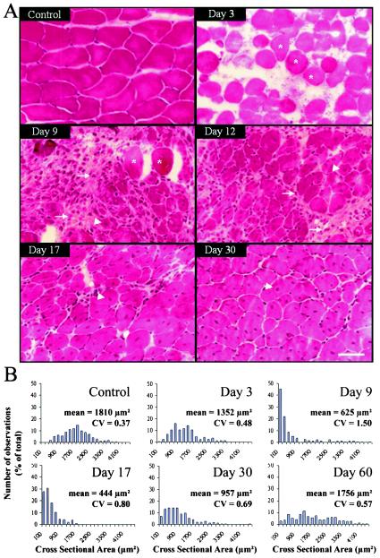
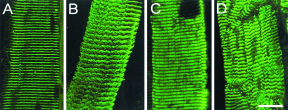
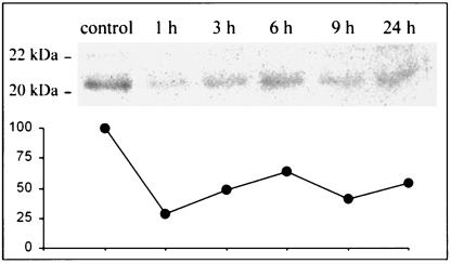
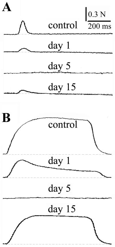
Clostridium sordellii lethal toxin (LT), a 250-kDa protein which is the bacteria's major virulence factor, belongs to a family of large clostridial cytotoxins which glucosylate small GTP-binding proteins. Here, we report the results of our ex vivo analysis of the structure and function of skeletal neuromuscular tissue obtained from mice at various times after intramuscular injection of a sublethal dose of LT (0.25 ng/g of body wt). The toxin caused, within 24 h, pronounced localized edema, inflammation, myofibril disassembly, and degeneration of skeletal muscle fibers in the injected area, and it glucosylated the muscle tissue's small GTPases. Regeneration of the damaged fibers was evident 6 to 9 days postinjury and was completed by 60 days. The expression of dystrophin, laminin, and fast and neonatal myosin in regenerating fibers, detected by immunofluorescence microscopy, confirmed that LT does not impair the high regenerative capacity of murine skeletal muscle fibers. Functional studies revealed that LT affects muscle contractility and neuromuscular transmission. However, partial recovery of nerve-evoked muscle twitches and tetanic contractions was observed by day 15 postinjection, and extensive remodeling of the neuromuscular junction's nerve terminals and clusters of muscle acetylcholine receptors was still evident 30 days postinjection. In conclusion, to the best of our knowledge, this is the first report to characterize the degeneration and regeneration of skeletal neuromuscular tissue after in vivo exposure to a large clostridial cytotoxin. In addition, our data may provide an explanation for the severe neuromuscular alterations accompanying wound infections caused by C. sordellii.
Figures
Similar articles
-
Alpha1-syntrophin-deficient skeletal muscle exhibits hypertrophy and aberrant formation of neuromuscular junctions during regeneration.J Cell Biol. 2002 Sep 16;158(6):1097-107. doi: 10.1083/jcb.200204076. Epub 2002 Sep 9. J Cell Biol. 2002. PMID: 12221071 Free PMC article.
-
Acetylcholine receptors and nerve terminal distribution at the neuromuscular junction of long-term regenerated muscle fibers.J Neurocytol. 2005 Dec;34(6):387-96. doi: 10.1007/s11068-006-8725-1. Epub 2006 Aug 10. J Neurocytol. 2005. PMID: 16902760
-
[Pathological changes in neuromuscular junction during ischemia-reperfusion in rat skeletal muscle].Zhongguo Xiu Fu Chong Jian Wai Ke Za Zhi. 2006 Nov;20(11):1103-8. Zhongguo Xiu Fu Chong Jian Wai Ke Za Zhi. 2006. PMID: 17191578 Chinese.
-
Presynaptic neurotoxins with enzymatic activities.Handb Exp Pharmacol. 2008;(184):129-70. doi: 10.1007/978-3-540-74805-2_6. Handb Exp Pharmacol. 2008. PMID: 18064414 Review.
-
Large clostridial cytotoxins.Rev Physiol Biochem Pharmacol. 2004;152:23-47. doi: 10.1007/s10254-004-0033-5. Epub 2004 Sep 21. Rev Physiol Biochem Pharmacol. 2004. PMID: 15449191 Review.
Cited by
-
Past injurious exercise attenuates activation of primary calcium-dependent injury pathways in skeletal muscle during subsequent exercise.Physiol Rep. 2018 Mar;6(6):e13660. doi: 10.14814/phy2.13660. Physiol Rep. 2018. PMID: 29595913 Free PMC article.
-
Clostridium sordellii lethal toxin kills mice by inducing a major increase in lung vascular permeability.Am J Pathol. 2007 Mar;170(3):1003-17. doi: 10.2353/ajpath.2007.060583. Am J Pathol. 2007. PMID: 17322384 Free PMC article.
-
Regional adaptation of collagen in skeletal muscle to repeated bouts of strenuous eccentric exercise.Pflugers Arch. 2016 Sep;468(9):1565-72. doi: 10.1007/s00424-016-1860-3. Epub 2016 Jul 28. Pflugers Arch. 2016. PMID: 27469054
-
Foot infection by Clostridium sordellii: case report and review of 15 cases in France.J Clin Microbiol. 2015 Apr;53(4):1423-7. doi: 10.1128/JCM.03414-14. Epub 2015 Jan 21. J Clin Microbiol. 2015. PMID: 25609723 Free PMC article.
-
Deficient Skeletal Muscle Regeneration after Injury Induced by a Clostridium perfringens Strain Associated with Gas Gangrene.Infect Immun. 2019 Jul 23;87(8):e00200-19. doi: 10.1128/IAI.00200-19. Print 2019 Aug. Infect Immun. 2019. PMID: 31138614 Free PMC article.
References
-
- Anderson, J. E. 1998. Studies of the dynamics of skeletal muscle regeneration: the mouse came back. Biochem. Cell Biol. 76:13-26. - PubMed
-
- Angaut-Petit, D., J. Molgó, A. Connold, and L. Faille. 1987. The levator auris longus muscle of the mouse: a convenient preparation for studies of short- and long-term presynaptic effects of drugs or toxins. Neurosci. Lett. 82:83-88. - PubMed
-
- Boquet, P. 1999. Bacterial toxins inhibiting or activating small GTP-binding proteins. Ann. N. Y. Acad. Sci. 886:83-90. - PubMed
Publication types
MeSH terms
Substances
LinkOut - more resources
Full Text Sources
