. 2004 Jun;72(6):3171-8.
doi: 10.1128/IAI.72.6.3171-3178.2004.

Identification of acyloxyacyl hydrolase, a lipopolysaccharide-detoxifying enzyme, in the murine urinary tract

Affiliations

Identification of acyloxyacyl hydrolase, a lipopolysaccharide-detoxifying enzyme, in the murine urinary tract

J Amelia Feulner et al. Infect Immun. 2004 Jun.

Abstract

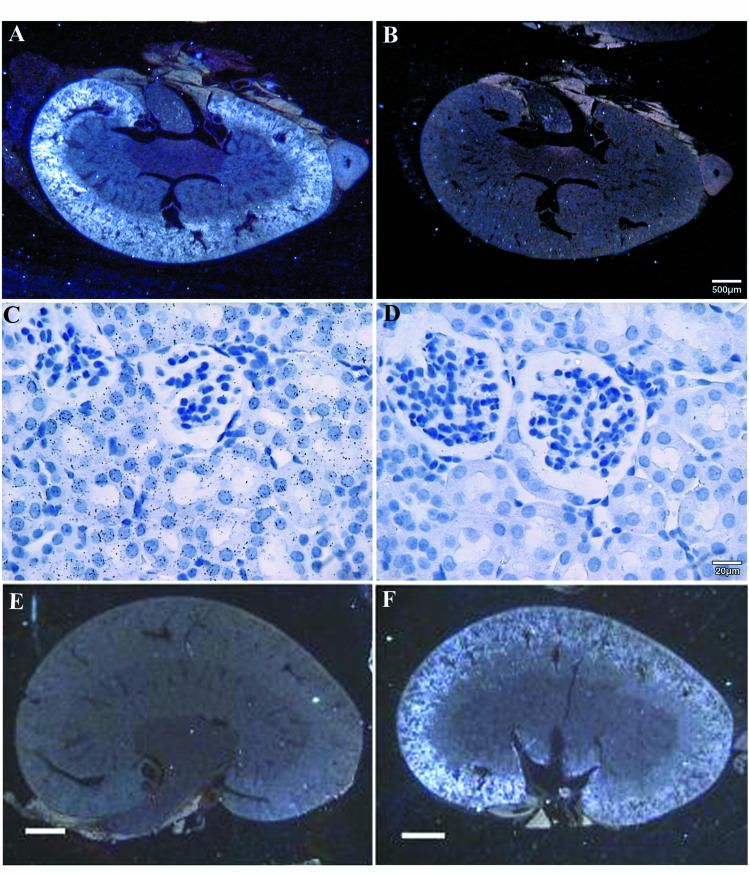
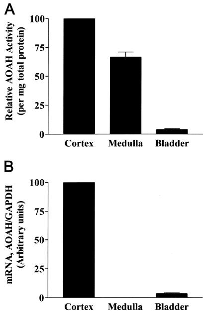
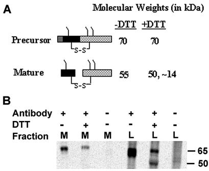
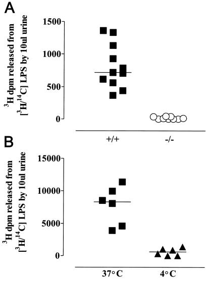
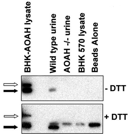
Acyloxyacyl hydrolase (AOAH) is an unusual but highly conserved lipase, previously described only in myeloid cells, that removes secondary fatty acyl chains from bacterial lipopolysaccharides (LPS) and may also act on various glycero(phospho)lipids. Deacylation by AOAH greatly reduces the ability of LPS to stimulate cells via CD14-MD-2-Toll-like receptor 4. We report here that renal cortical tubule cells produce AOAH and secrete it into urine, where it can deacylate LPS. In vitro studies revealed that proximal tubule cells secrete pro-AOAH, which can be taken up by bladder cells and processed to the heterodimeric, more enzymatically active, mature form of AOAH. AOAH can then be used by the recipient cells to deacylate LPS. The enzyme produced by proximal tubule epithelium may thus be shared with downstream cells. In addition, mature AOAH is found in the urine. We suggest that cortical tubule cells may produce and secrete AOAH to limit inflammatory responses to gram-negative bacteria throughout the urinary tract.


Figures

FIG. 1.
FIG. 1.
AOAH expression in murine tissues. (A) Northern analysis of multiple tissue RNAs from C57BL/6 mice (Clontech) with labeled AOAH cDNA as probe (top). The blot was stripped and reprobed to detect β-actin (bottom). The migration positions of size markers (in kilobase pairs) are shown. (B) AOAH activity in lysates of freshly harvested C57BL/6 mouse tissues. Measurements were performed in duplicate. The bars show means plus one standard deviation of data from three independent experiments.
FIG. 2.
FIG. 2.
Localization of AOAH mRNA in murine kidney by in situ hybridization. Panels: A, C, E, and F, antisense probe; B and D, sense probe. Panels A to D are from ICR (outbred) mice, whereas panels E and F are from AOAH −/− (E) and +/+ (F) 129 mice. The antisense probe hybridized to the renal cortex (panels A and F) and was found over cortical tubules but not glomeruli (panel C). The bars indicate 500 μm (A and B), 20 μm (C and D), or 1 mm (E and F). The experiment shown in panels A to D was repeated three times; each experiment used kidney from a different mouse. The results shown in panels E and F were confirmed with kidneys from AOAH +/+ and AOAH −/− C57BL/6 mice.
FIG. 3.
FIG. 3.
AOAH is synthesized in the renal cortex. (A) AOAH activity in lysates of renal cortex, medulla, and washed bladder. Each bar shows the mean and standard error of three or more measurements. The mean activity in seven bladder samples was 2.5% of the activity in total kidney lysates. (B) Real-time PCR analysis of AOAH and GAPDH mRNA in renal cortex, medulla, and bladder. This experiment was performed in duplicate and repeated twice, using tissues from different mice, with similar results.
FIG. 4.
FIG. 4.
Proximal tubule cells secrete pro-AOAH. (A) Diagram of AOAH structure, showing the conversion of the precursor (pro-AOAH) into mature AOAH. (B) Production of 35S-AOAH by porcine proximal tubule cells in vitro. Labeled AOAH was immunoprecipitated as described in Methods and studied by SDS-PAGE and autoradiography. M, medium; L, lysate. Antibody +, anti-AOAH; antibody −, preimmune IgG. Only pro-AOAH is seen in the media, whereas the lysate contains both pro-AOAH and mature AOAH.
FIG. 5.
FIG. 5.
AOAH activity in mouse urine. (A) LPS deacylation by urine from AOAH +/+ and −/− mice. Urine (10 μl) was added to 490 μl of AOAH reaction mixture containing 1 μg of [3H/14C]LPS and incubated at 37°C for 18 h before the addition of ethanol and further steps as described in Materials and Methods. (B) LPS deacylation by murine urine. Fresh urine (10 μl) was incubated with [3H/14C]LPS (0.5 μg) at 4 and 37°C for 18 h. AOAH reaction mixture was then added to provide protein for coprecipitation of intact LPS, followed by ethanol. The remaining steps are described in Materials and Methods. Each datum point represents a different mouse.
FIG. 6.
FIG. 6.
Mature AOAH is found in urine. Equal volumes of wild-type and AOAH-null urine were immunoprecipitated with an anti-murine AOAH monoclonal antibody and assayed by Western blot as described in Materials and Methods. Lysates of BHK cells transfected with AOAH were used as the positive control. The results are representative of three experiments with similar results. Note in the upper panel that the BHK-AOAH cell lysate contains both pro-AOAH (open arrow) and mature AOAH (solid arrow). Wild-type urine only has mature AOAH. After treatment with DTT (lower panel), mature AOAH migrates with an apparent Mr of 50 kDa (solid arrow). The band at an apparent Mr of 55 kDa in the lower panel is the heavy chain of the murine monoclonal antibody used for immunoprecipitation.
FIG. 7.
FIG. 7.
Bladder cells take up AOAH precursor. (A) Uptake of pro-AOAH by T24 cells after incubation with pro-AOAH-containing medium for 5 h in the presence or absence of 10 mM M6P, G6P, or mannose. Washed cells were lysed and AOAH protein was assayed by ELISA. Each bar shows the mean and standard error of data from four independent experiments. (B) AOAH specific activity (activity/nanograms of protein) in T24 cell lysates. Compared to the pro-AOAH added in the medium (AOAH Source), cell-associated AOAH had a much greater specific activity, reflecting its activation by the T24 cells. M6P inhibited AOAH binding (A) but did not prevent the activation of the cell-associated AOAH (B).
FIG. 8.
FIG. 8.
AOAH confers LPS-deacylating activity to bladder cells. T24 cells were allowed to take up AOAH for 5 h, washed, and then incubated with [3H]LPS (125 ng/ml) for the times indicated. Control cells were incubated with medium that did not contain AOAH. Whereas control and AOAH-containing cells took up similar amounts of [3H]LPS (A), only the AOAH-containing cells removed 3H-labeled fatty acids from the LPS backbone (B). The data represent combined results of three separate experiments; the error bars represent one standard error of the mean. Solid squares and bars, T24 cells with AOAH; open circles and bars, control T24 cells.

References

    1. Bäckhed, F., M. Söderhäll, P. Ekmann, S. Normark, and A. Richter-Dahlfors. 2001. Induction of innate immune responses by Escherichia coli and purified lipopolysaccharide correlate with organ- and cell-specific expression of Toll-like receptors within the human urinary tract. Cell. Microbiol. 3:153-158. - PubMed
    1. Chippendale, G. R., J. W. Warren, A. L. Trifillis, and H. L. T. Mobley. 1994. Internalization of Proteus mirabilis by human renal epithelial cells. Infect. Immun. 62:3115-3121. - PMC - PubMed
    1. Coulthard, M. G., J. Swindle, R. S. Munford, R. D. Gerard, and R. S. Meidell. 1996. Adenovirus-mediated transfer of a gene encoding acyloxyacyl hydrolase (AOAH) into mice increases tissue and plasma AOAH activity. Infect. Immun. 64:1510-1515. - PMC - PubMed
    1. Donnenberg, M. S., B. Newman, S. J. Utsalo, A. L. Trifillis, J. R. Hebel, and J. W. Warren. 1994. Internalization of Escherichia coli into human kidney epithelial cells: comparison of fecal and pyelonephritis-associated strains. J. Infect. Dis. 169:831-838. - PubMed
    1. Guo, L., K. B. Lim, J. S. Gunn, B. Bainbridge, R. P. Darveau, M. Hackett, and S. I. Miller. 1997. Regulation of lipid A modifications by Salmonella typhimurium virulence genes phoP-phoQ. Science 276:250-253. - PubMed

Publication types