. 2004 Jun;72(6):3299-309.
doi: 10.1128/IAI.72.6.3299-3309.2004.

Bacterial probiotic modulation of dendritic cells

Affiliations

Bacterial probiotic modulation of dendritic cells

Maureen Drakes et al. Infect Immun. 2004 Jun.

Abstract

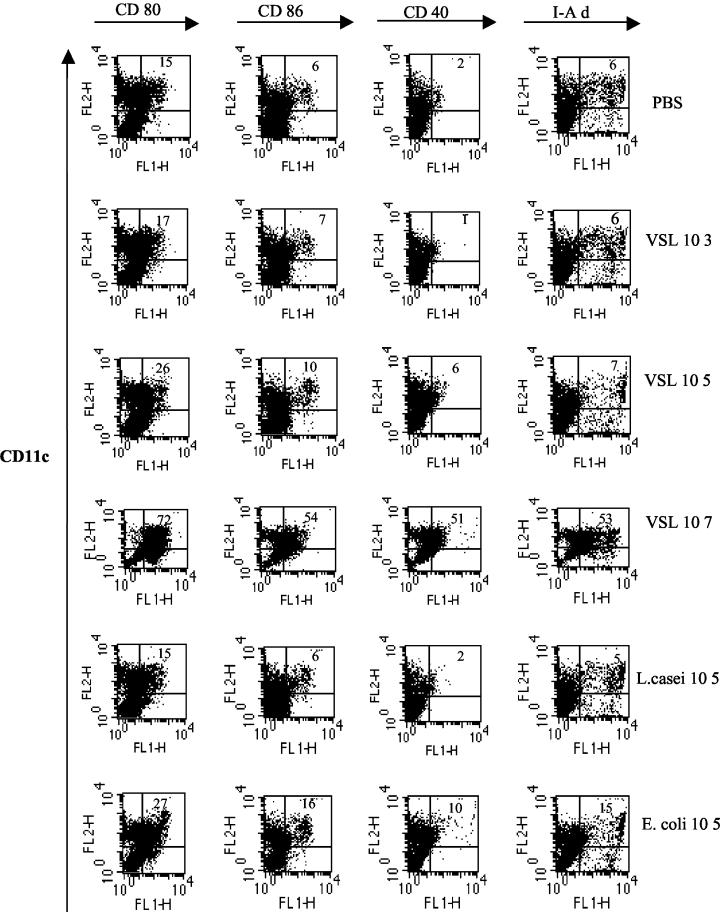
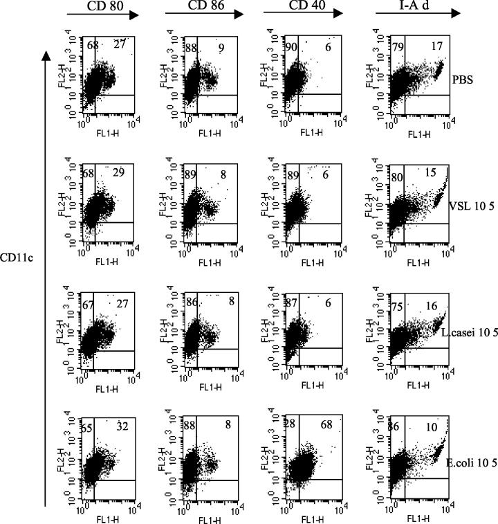
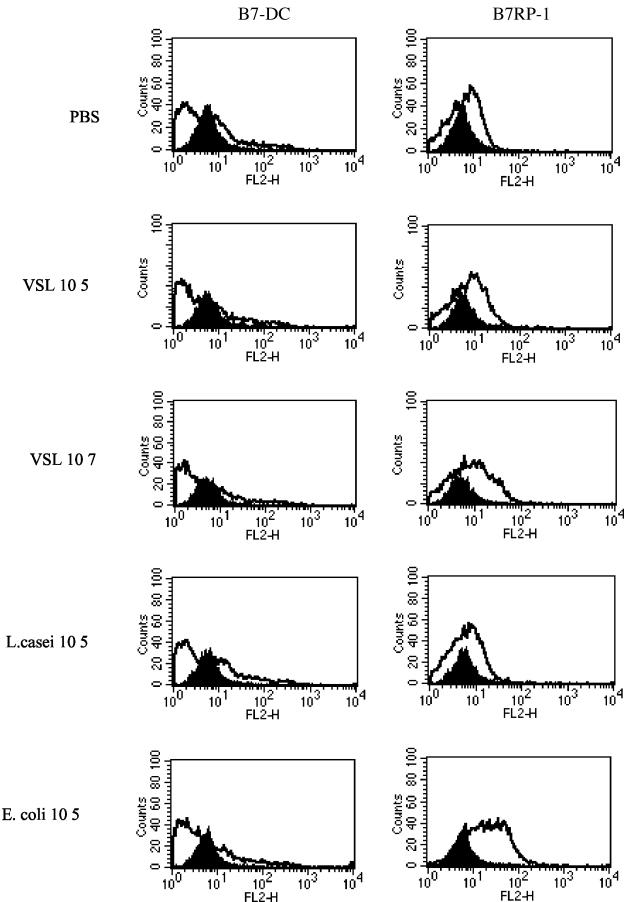
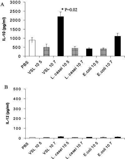
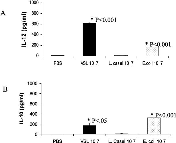
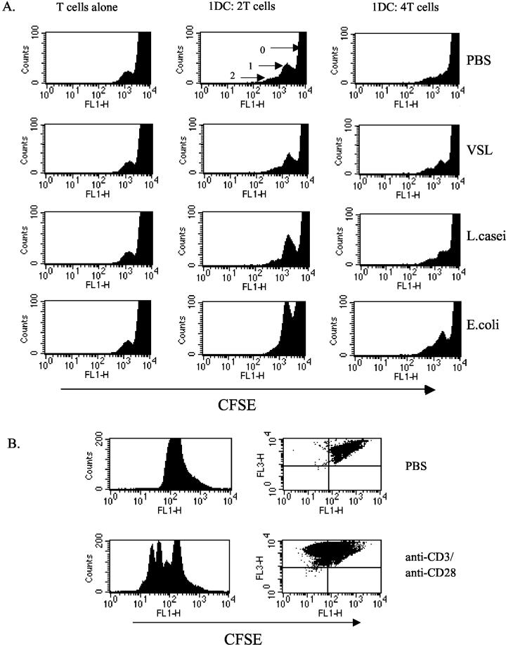
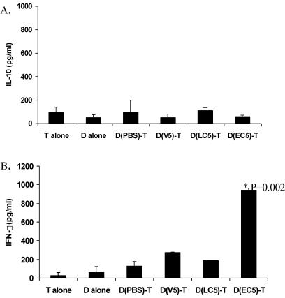
Intestinal dendritic cells are continually exposed to ingested microorganisms and high concentrations of endogenous bacterial flora. These cells can be activated by infectious agents and other stimuli to induce T-cell responses and to produce chemokines which recruit other cells to the local environment. Bacterial probiotics are of increasing use against intestinal disorders such as inflammatory bowel disease. They act as nonpathogenic stimuli within the gut to regain immunologic quiescence. This study was designed to determine the ability of a bacterial probiotic cocktail VSL#3 to alter cell surface antigen expression and cytokine production in bone marrow-derived dendritic cell-enriched populations. Cell surface phenotype was monitored by monoclonal fluorescent antibody staining, and cytokine levels were quantitated by enzyme-linked immunosorbent assay. High-dose probiotic upregulated the expression of C80, CD86, CD40, and major histocompatibility complex class II I-Ad. Neither B7-DC or B7RP-1 was augmented after low-dose probiotic or Lactobacillus casei treatment, but B7RP-1 showed increased expression on dendritic cells stimulated with the gram-negative bacterium Escherichia coli. Functional studies showed that probiotic did not enhance the ability of dendritic cells to induce allogeneic T-cell proliferation, as was observed for E. coli. Substantial enhancement of interleukin-10 release was observed in dendritic cell-enriched culture supernatants after 3 days of probiotic stimulation. These results demonstrate that probiotics possess the ability to modulate dendritic cell surface phenotype and cytokine release in granulocyte-macrophage colony-stimulating factor-stimulated bone marrow-derived dendritic cells. Regulation of dendritic cell cytokines by probiotics may contribute to the benefit of these molecules in treatment of intestinal diseases.


Figures

FIG. 1.
FIG. 1.
Regulation of DC surface phenotypes by probiotic. BM cells were grown in GM-CSF for 2 days. Stimulation as indicated was added on day 2 for a further 3 days. Cells were harvested and MAb double stained for CD11c PE (FL2)- and FITC (FL1)-labeled costimulatory molecules. Gating was done to exclude dead cells. The number shown in the top right hand corner of each dot plot quadrant represents the percentage of CD11c-positive cells expressing each marker as indicated. Probiotic VSL#3 is indicated as VSL in the figures. Results are representative of two separate experiments.
FIG. 2.
FIG. 2.
Low-dose probiotic maintains immature DC cell surface phenotype. DC cultures were harvested on day 5. Cells were treated with probiotic or bacteria overnight, and cell surface phenotypes were characterized by staining for CD11c PE- and FITC-labeled costimulatory molecules. Dead cells were gated out, and 10,000 events were collected on a FACScan. The percentage of double-positive cells is shown in the upper right quadrant, and the percentage of CD11c single-positive cells is shown in the upper left quadrant. The results shown are representative of three separate experiments.
FIG. 3.
FIG. 3.
Bacteria differentially modulate B7RP-1 expression on DC. (A) DC cultures were harvested on day 5, further enriched on a CD11c MACS column, and cocultured with probiotic, L. casei, or E. coli at 105 organisms/ml for an overnight period. Cells were double stained for B7-DC or B7RP-1 PE and CD11c FITC. The solid histogram shows results for immunoglobulin G controls, and the unshaded histogram area shows the level of expression of costimulatory molecule as a result of treatment. Results for upregulation of B7RP-1 by E. coli are representative of three separate experiments.
FIG. 4.
FIG. 4.
Probiotic stimulates IL-10 release in DC cultures. BM cells were grown in GM-CSF. Various concentrations of probiotic or bacteria were added on day 2 for a further 3 days. DC culture supernatants were harvested on day 5, and cytokine levels were measured by ELISA. Results are shown for IL-10 (A) and IL-12 (p70) (B). Error bars represent standard deviations from duplicate wells. P values indicate levels of significance above control PBS. Results are representative of three separate experiments.
FIG. 5.
FIG. 5.
Probiotic stimulation of IL-12 (p70). Cells at day 4 of BM DC culture were stimulated for an overnight period by the addition of 107 organisms of probiotic or bacteria/ml until the next day. Supernatants were collected, and IL-12 levels (A) or IL-10 release (B) in supernatants were determined by ELISA. Standard deviation is shown for duplicate wells. The level of significance is indicated by the P value.
FIG. 6.
FIG. 6.
Probiotic-stimulated DC does not enhance T-cell division in allogeneic T cells. (A) Day 5 BM-derived DC cultures were harvested, and CD11c cells were purified and treated (2 × 10⁶ DC/ml in 24-well plates) with probiotic or bacteria (105 organisms per ml) for an overnight culture period. DC were washed three times. CD4 T cells were purified from the spleens of allogeneic C57BL6/J mice and labeled with 0.5 μM CFSE. Serial dilutions were made of DC which were then added to 96-well flat-bottom plates at the indicated dilution, with 2 × 10⁵ T cells/well. After 4 days, cells were harvested and MAb labeled for CD4 PerCP (FL3). Forward scatter versus side scatter gating was done on live cells, and 50,000 events were collected. Histograms represent the CD4+ PerCP-stained T cells which contained the CFSE dye (FL1). The peak labeled 0 shows the undivided cells, the peak labeled 1 shows the first round of cell division, and the peak labeled 2 shows those cells undergoing a second division. Probiotic-stimulated DC induced minimal T-cell division above control PBS in three separate experiments. (B) Plates were coated with 1-μg/ml anti-CD3 MAb in PBS at 37°C for 4 h. Wells were washed gently with PBS. CFSE-labeled T cells (2 × 10⁶/ml) were added in RPMI medium with supplements, and 2-μg/ml soluble anti-CD28 was added to the cultures. Cells were harvested at day 3 of stimulation and stained with CD4 PerCP, and the division of cells was analyzed as described above. Dot plots show divided cells in the upper left quadrant.
FIG. 7.
FIG. 7.
Cytokine production in DC and T-cell cocultures. Day 5 BM-derived DC generated from BALB/c mice were harvested, purified for CD11c-positive cells, and treated with probiotic or bacteria (105 organisms/ml). Treated DC-purified T-cell cocultures were set up with 0.5 × 10⁶ DC and 1.5 × 10⁶ T cells in 24-well plates. Supernatants were harvested at day 3 and frozen at −20°C for cytokine analysis by ELISA. D alone represents DC precultured in PBS; similar results were obtained with those precultured in bacteria or probiotic. D(V5), DC pretreated with VSL#3 at 105 organisms/ml; D(LC5), pretreated with L. casei; D(EC5), those pretreated with E. coli. Values represent means ± 1 standard deviation of the results for duplicate wells from two experiments performed. (A) IL-10 production; (B) IFN-γ production in cocultures.

References

    1. Alakomi, H. L., E. Skytta, M. Saarela, T. Mattila-Sandholm, K. Latva-Kala, and I. M. Helander. 2000. Lactic acid permeabilizes gram-negative bacteria by disrupting the outer membrane. Appl. Environ. Microbiol. 66:2001-2005. - PMC - PubMed
    1. Alander, M., R. Satokari, R. Korpela, M. Saxelin, T. Vilpponen-Salmela, T. Mattila-Sandholm, and A. von Wright. 1999. Persistence of colonization of human colonic mucosa by a probiotic strain, Lactobacillus rhamnosus GG, after oral consumption. Appl. Environ. Microbiol. 65:351-354. - PMC - PubMed
    1. Annuk, H., J. Shchepetova, T. Kullisaar, E. Songisepp, M. Zilmer, and M. Mikelsaar. 2003. Characterization of intestinal lactobacilli as putative probiotic candidates. J. Appl. Microbiol. 94:403-412. - PubMed
    1. Asseman, C., S. Mauze, M. W. Leach, R. L. Coffman, and F. Powrie. 1999. An essential role for interleukin 10 in the function of regulatory T cells that inhibit intestinal inflammation. J. Exp. Med. 190:995-1004. - PMC - PubMed
    1. Banchereau, J., and R. M. Steinman. 1998. Dendritic cells and the control of immunity. Nature 392:245-252. - PubMed

Publication types