

Cryptococcus neoformans capsule structure evolution in vitro and during murine infection
- PMID: 15155641
- PMCID: PMC415706
- DOI: 10.1128/IAI.72.6.3359-3365.2004
Cryptococcus neoformans capsule structure evolution in vitro and during murine infection
Abstract
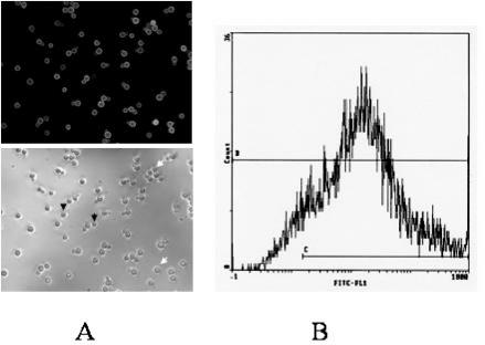
Cryptococcus neoformans capsule structure modifications after prolonged in vitro growth or in vivo passaging have been reported previously. However, nothing is known about the dynamics of these modifications or about their environmental specificities. In this study, capsule structure modifications after mouse passaging and prolonged in vitro culturing were analyzed by flow cytometry using the glucuronoxylomannan-specific monoclonal antibody E1. The capsule structures of strains recovered after 0, 1, 8, and 35 days were compared by using the level of E1-specific epitope expression and its cell-to-cell heterogeneity within a given cell population. In vitro, according to these parameters, the diversity of the strains was higher on day 35 than it was initially, suggesting the absence of selection during in vitro culturing. In contrast, the diversity of the strains recovered from the brain tended to decrease over time, suggesting that selection of more adapted strains had occurred. The strains recovered on day 35 from the spleen and the lungs had different phenotypes than the strains isolated from the brain of the same mouse on the same day, thus strongly suggesting that there is organ specificity for C. neoformans strain selection. Fingerprinting of the strains recovered in vitro and in vivo over time confirmed that genotypes evolved very differently in vitro and in vivo, depending on the environment. Overall, our results suggest that organ-specific selection can occur during cryptococcosis.
Figures


Similar articles
-
Anti-glucuronoxylomannan IgG1 specific antibodies production in Cryptococcus neoformans resistant mice.Biomedica. 2005 Mar;25(1):110-9. Biomedica. 2005. PMID: 15962907
-
An anti-beta-glucan monoclonal antibody inhibits growth and capsule formation of Cryptococcus neoformans in vitro and exerts therapeutic, anticryptococcal activity in vivo.Infect Immun. 2007 Nov;75(11):5085-94. doi: 10.1128/IAI.00278-07. Epub 2007 Jul 2. Infect Immun. 2007. PMID: 17606600 Free PMC article.
-
Systematic capsule gene disruption reveals the central role of galactose metabolism on Cryptococcus neoformans virulence.Mol Microbiol. 2007 May;64(3):771-81. doi: 10.1111/j.1365-2958.2007.05695.x. Mol Microbiol. 2007. PMID: 17462022
-
Deciphering the model pathogenic fungus Cryptococcus neoformans.Nat Rev Microbiol. 2005 Oct;3(10):753-64. doi: 10.1038/nrmicro1245. Nat Rev Microbiol. 2005. PMID: 16132036 Review.
-
Cryptococcus neoformans: a sugar-coated killer with designer genes.FEMS Immunol Med Microbiol. 2005 Sep 1;45(3):395-404. doi: 10.1016/j.femsim.2005.06.005. FEMS Immunol Med Microbiol. 2005. PMID: 16055314 Review.
Cited by
-
Size Matters: Measurement of Capsule Diameter in Cryptococcus neoformans.J Vis Exp. 2018 Feb 27;(132):57171. doi: 10.3791/57171. J Vis Exp. 2018. PMID: 29553511 Free PMC article.
-
Mixed infections and In Vivo evolution in the human fungal pathogen Cryptococcus neoformans.mBio. 2010 May 18;1(1):e00091-10. doi: 10.1128/mBio.00091-10. mBio. 2010. PMID: 20689742 Free PMC article.
-
Cryptococcus neoformans induces antimicrobial responses and behaves as a facultative intracellular pathogen in the non mammalian model Galleria mellonella.Virulence. 2015;6(1):66-74. doi: 10.4161/21505594.2014.986412. Virulence. 2015. PMID: 25531532 Free PMC article.
-
Radial mass density, charge, and epitope distribution in the Cryptococcus neoformans capsule.Eukaryot Cell. 2007 Jan;6(1):95-109. doi: 10.1128/EC.00306-06. Epub 2006 Nov 17. Eukaryot Cell. 2007. PMID: 17114596 Free PMC article.
-
Antigenic and phenotypic variations in fungi.Cell Microbiol. 2009 Dec;11(12):1716-23. doi: 10.1111/j.1462-5822.2009.01384.x. Epub 2009 Sep 21. Cell Microbiol. 2009. PMID: 19769677 Free PMC article. Review.
References
-
- Brandt, M. E., C. Hutwagner, L. A. Klug, W. S. Baughman, D. Rimland, E. A. Graviss, R. J. Hamill, C. Thomas, P. G. Pappas, A. L. Reingold, R. W. Pinner, and the Cryptococcal Disease Active Surveillance Group. 1996. Molecular subtype distribution of Cryptococcus neoformans in four areas of the United States. J. Clin. Microbiol. 34:912-917. - PMC - PubMed
Publication types
MeSH terms
Substances
LinkOut - more resources
Full Text Sources
Other Literature Sources
