

Role of CD8+ and CD4+ T lymphocytes in jejunal mucosal injury during murine giardiasis
- PMID: 15155662
- PMCID: PMC415705
- DOI: 10.1128/IAI.72.6.3536-3542.2004
Role of CD8+ and CD4+ T lymphocytes in jejunal mucosal injury during murine giardiasis
Abstract
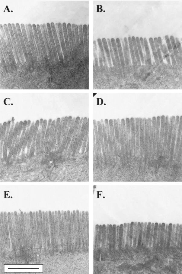
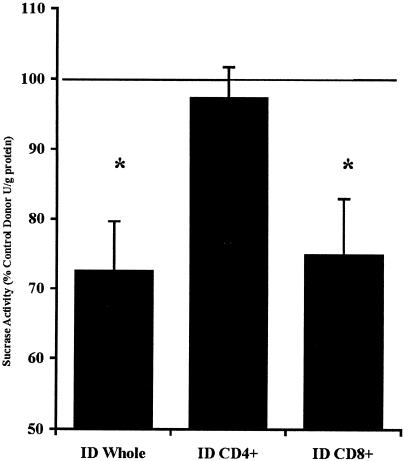
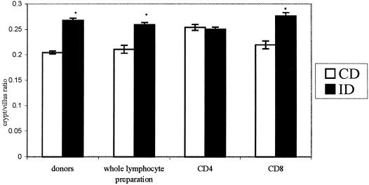
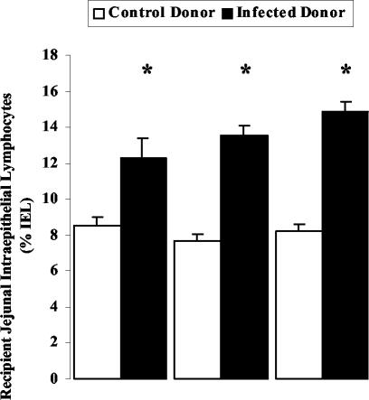
T-cell-mediated pathogenesis has been documented in various idiopathic and microbially induced intestinal disorders. Diffuse microvillous shortening seen in giardiasis is responsible for disaccharidase insufficiencies and malabsorption of electrolytes, nutrients, and water. Other mucosal changes include crypt hyperplasia and increased numbers of intraepithelial lymphocytes (IEL). A recent report using an athymic mouse model of infection showed that these epithelial injuries were dependent on T cells. The aim of the present study was to identify which subset of superior mesenteric lymph node (SMLN) T cells were responsible for mucosal alterations in giardiasis. CD4+ and CD8+ T cells, as well as whole lymphocyte populations, were isolated from SMLN of Giardia muris-infected mice for adoptive transfer. Jejunal segments of recipient mice were assessed for brush border ultrastructure, sucrase activity, crypt/villus ratio, and IEL numbers. Mice that received enriched CD8+ and whole SMLN lymphocytes, but not CD4+ T cells, from infected donors showed diffuse shortening of microvilli, loss of brush border surface area, impaired sucrase activity, and increased crypt/villus ratios compared to respective controls. Transfer of whole SMLN lymphocytes, as well as enriched CD4+ or CD8+ T cells, from infected donors led to increased IEL numbers in the recipient jejunum. The findings indicate that loss of intestinal brush border surface area, reduced disaccharidase activities, and increased crypt/villus ratios in giardiasis are mediated by CD8+ T cells, whereas both CD8+ and CD4+ SMLN T cells regulate the influx of IEL.
Figures
Similar articles
-
The Microbiota Contributes to CD8+ T Cell Activation and Nutrient Malabsorption following Intestinal Infection with Giardia duodenalis.Infect Immun. 2016 Sep 19;84(10):2853-60. doi: 10.1128/IAI.00348-16. Print 2016 Oct. Infect Immun. 2016. PMID: 27456829 Free PMC article.
-
Jejunal brush border microvillous alterations in Giardia muris-infected mice: role of T lymphocytes and interleukin-6.Infect Immun. 2000 Jun;68(6):3412-8. doi: 10.1128/IAI.68.6.3412-3418.2000. Infect Immun. 2000. PMID: 10816492 Free PMC article.
-
Immunopathology of giardiasis: the role of lymphocytes in intestinal epithelial injury and malfunction.Mem Inst Oswaldo Cruz. 2005 Mar;100 Suppl 1:185-90. doi: 10.1590/s0074-02762005000900032. Epub 2005 Jun 14. Mem Inst Oswaldo Cruz. 2005. PMID: 15962121 Review.
-
Effects of murine giardiasis on growth, intestinal morphology , and disaccharidase activity.J Parasitol. 1990 Jun;76(3):403-9. J Parasitol. 1990. PMID: 2191103
-
Recent insights into the mucosal reactions associated with Giardia lamblia infections.Int J Parasitol. 2005 Nov;35(13):1339-47. doi: 10.1016/j.ijpara.2005.07.008. Epub 2005 Aug 24. Int J Parasitol. 2005. PMID: 16182298 Review.
Cited by
-
Intestinal immune responses to commensal and pathogenic protozoa.Front Immunol. 2022 Sep 16;13:963723. doi: 10.3389/fimmu.2022.963723. eCollection 2022. Front Immunol. 2022. PMID: 36211380 Free PMC article. Review.
-
Inflammation drives dysbiosis and bacterial invasion in murine models of ileal Crohn's disease.PLoS One. 2012;7(7):e41594. doi: 10.1371/journal.pone.0041594. Epub 2012 Jul 25. PLoS One. 2012. PMID: 22848538 Free PMC article.
-
The Microbiota Contributes to CD8+ T Cell Activation and Nutrient Malabsorption following Intestinal Infection with Giardia duodenalis.Infect Immun. 2016 Sep 19;84(10):2853-60. doi: 10.1128/IAI.00348-16. Print 2016 Oct. Infect Immun. 2016. PMID: 27456829 Free PMC article.
-
Impaired Intestinal Barrier and Tissue Bacteria: Pathomechanisms for Metabolic Diseases.Front Endocrinol (Lausanne). 2021 Mar 9;12:616506. doi: 10.3389/fendo.2021.616506. eCollection 2021. Front Endocrinol (Lausanne). 2021. PMID: 33767669 Free PMC article. Review.
-
Interactions between parasites and microbial communities in the human gut.Front Cell Infect Microbiol. 2012 Nov 16;2:141. doi: 10.3389/fcimb.2012.00141. eCollection 2012. Front Cell Infect Microbiol. 2012. PMID: 23162802 Free PMC article. Review.
References
-
- Adjei, A. A., J. T. Jones, and F. J. Enriquez. 2000. Differential intra-epithelial lymphocyte phenotypes following Cryptosporidium parvum challenge in susceptible and resistant athymic strains of mice. Parasitol. Int. 49:119-129. - PubMed
-
- Arvanitakis, C. 1979. Abnormalities of jejunal mucosal enzymes in ulcerative colitis and Crohn's disease. Digestion 19:259-266. - PubMed
-
- Buret, A., D. G. Gall, and M. E. Olson. 1990. Effects of murine giardiasis on growth, intestinal morphology, and disaccharidase activity. J. Parasitol. 76:403-409. - PubMed
-
- Buret, A., D. G. Gall, and M. E. Olson. 1991. Growth, activities of enzymes in the small intestine, and ultrastructure of microvillous border in gerbils infected with Giardia duodenalis. Parasitol. Res. 77:109-114. - PubMed
Publication types
MeSH terms
Substances
LinkOut - more resources
Full Text Sources
Other Literature Sources
Medical
Research Materials
