. 2004 Jun;72(6):3571-6.
doi: 10.1128/IAI.72.6.3571-3576.2004.

Infection with Toxoplasma gondii increases atherosclerotic lesion in ApoE-deficient mice

Affiliations

Infection with Toxoplasma gondii increases atherosclerotic lesion in ApoE-deficient mice

Luciane R Portugal et al. Infect Immun. 2004 Jun.

Abstract

Toxoplasma gondii is an intracellular protozoan that elicits a potent inflammatory response during the acute phase of infection. Herein, we evaluate whether T. gondii infection alters the natural course of aortic lesions. ApoE knockout mice were infected with T. gondii, and at 5 weeks of infection, serum, feces, and liver cholesterol; aortic lesion size, cellularity, and inflammatory cytokines; and levels of serum nitrite and gamma interferon (IFN-gamma) were analyzed. Our results showed that serum cholesterol and atherogenic lipoproteins were reduced after T. gondii infection. The reduction of serum levels of total cholesterol and atherogenic lipoproteins was associated with increases in the aortic lesion area, numbers of inflammatory cells, and expression of monocyte chemoattractant protein 1 and inducible nitric oxide synthase mRNA in the site of lesions as well as elevated concentrations of IFN-gamma and nitrite in sera of T. gondii-infected animals. These results suggest that infection with T. gondii accelerates atherosclerotic development by stimulating the proinflammatory response and oxidative stress, thereby increasing the area of aortic lesion.


Figures

FIG. 1.
FIG. 1.
(A) Total serum cholesterol of noninfected (n = 14, black bars) or T. gondii-infected ApoE KO mice (n = 17, gray bars) after 0, 4, and 5 weeks postinfection. (B) Lipoprotein profile of noninfected (n = 5, fine line) and T. gondii-infected (n = 5, bold line) ApoE KO mice after 5 weeks of experiment (VLDL, fractions 5 to 10; IDL + LDL, fractions 11 to 19; HDL, fractions 21 to 26). The averages ± standard errors of the VLDL, IDL + LDL, and HDL fractions were 355 ± 51, 99 ± 15, and 1.2 ± 15 for the control group and 175 ± 14, 109 ± 7, and 5.2 ± 2.1 for the T. gondii group, respectively. (C) IFN-γ concentration in serum of noninfected (n = 6, black bars) or T. gondii-infected ApoE KO mice (n = 6, gray bars) at the beginning of and during the experiment. (D) Serum nitrite/nitrate concentration (micromoles/liter) of noninfected (n = 9, black bar) and T. gondii-infected (n = 12, gray bar) ApoE KO mice after 5 weeks of experiment. *P < 0.05.
FIG. 2.
FIG. 2.
Aorta measurement of the lesion area expressed as 1,000 micrometer2 (n = 11 noninfected mice and 12 T. gondii-infected mice) (A), inflammatory infiltrate (n = 6 per group); phenotype of cells from control group (B), phenotype of inflammatory cells (n = 6 per group) (C), and percentage of collagen (n = 6 per group) (D) in noninfected and T. gondii-infected ApoE KO mice after 5 weeks of experiment. Squares (A), diamonds (C), and circles (D) represent individual measurements. Horizontal lines represent the averages of groups. (B) Hatched and black bars represent the average of number of cells per 100 fields in 7 infected and 3 control animals, respectively. *, P < 0.05.
FIG. 3.
FIG. 3.
Histological aspect of the aorta of noninfected and T. gondii-infected ApoE KO mice after 5 weeks of experiment; typical lesions of control (A) and infected (B) mice are within the dashed square. (B) Presence of inflammatory focus (star) in Toxoplasma-infected group but not in control group. (C) Presence of well-defined fatty streak in control group, constituted by xantomized macrophages (arrow). (D) In T. gondii-infected animals, the lesions are more developed, with areas of cellular proliferation (arrow) and degenerative alterations (star), characterizing a more advanced stage of lesion. (E and F) Presence of cyst (within dashed square) in cardiac muscle near the aortic valve insertion (star). Note that there is no infiltration around the cyst. (CD4, CD8, and F4/80) Immunohistochemistry of atherosclerosis lesions in Toxoplasma-infected animals. Arrows in indicate cells stained with CD4-, CD8-, and F4/80-specific monoclonal antibodies, respectively. Bars, 200 μm (A and B), 35 μm (C and D), 70 μm (E), 11 μm (F), and 25 μm (CD4, CD8, and F4/80).
FIG. 4.
FIG. 4.
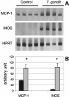
(A) Expression of the MCP-1 and iNOS endogenous mRNA in aortic tissue of noninfected (n = 4) and T. gondii-infected (n = 4) ApoE KO mice. (B) Densitometric analysis of control (black bar) and T. gondii-infected (gray bar) bands. The quantification was normalized to the levels of HPRT expression. Bars represent means and vertical lines represent standard errors for each group. *, statistical difference (P < 0.05) compared to the noninfected group.

Similar articles

Cited by

References

    1. Aliberti, J. C. S., F. S. Machado, J. T. Souto, A. P. Campanelli, M. M. Teixeira, R. T. Gazzinelli, and J. S. Silva. 1999. β-Chemokines enhance parasite uptake and promote nitric oxide-dependent microbiostatic activity in murine inflammatory macrophages infected with Trypanosoma cruzi. Infect. Immun. 67:4819-4826. - PMC - PubMed
    1. Binder, C. J., M. Chang, P. X. Shaw, Y. I. Miller, K. Hartvigsen, A. Dewan, and J. L. Witztum. 2002. Innate and acquired immunity in atherogenesis. Nat. Med. 8:1218-1226. - PubMed
    1. Blader, I. J., I. D. Manger, and J. C. Boothroyd. 2001. Microarray analysis reveals previously unknown changes in Toxoplasma gondii-infected human cells. J. Biol. Chem. 276:24223-27231. - PubMed
    1. Camargo, M. M., I. C. Almeida, M. E. S. Pereira, M. A. J. Ferguson, L. R. Travassos, and R. T. Gazzinelli. 1997. Glycosylphosphatidylinositol anchored mucin-like glycoproteins isolated from Trypanosoma cruzi trypomastigotes initiate the synthesis of pro-inflammatory cytokines by macrophages. J. Immunol. 158:5890-5901. - PubMed
    1. Coppens, I., A. P. Sinai, and K. A. Joiner. 2000. Toxoplasma gondii exploits host low-density lipoprotein receptor-mediated endocytosis for cholesterol acquisition. J. Cell Biol. 149:167-180. - PMC - PubMed

Publication types

MeSH terms

LinkOut - more resources