. 2004 Jun;16(6):1433-45.
doi: 10.1105/tpc.021568. Epub 2004 May 21.

Constitutive photomorphogenesis 1 and multiple photoreceptors control degradation of phytochrome interacting factor 3, a transcription factor required for light signaling in Arabidopsis

Affiliations

Constitutive photomorphogenesis 1 and multiple photoreceptors control degradation of phytochrome interacting factor 3, a transcription factor required for light signaling in Arabidopsis

Diana Bauer et al. Plant Cell. 2004 Jun.

Abstract

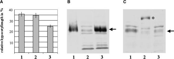
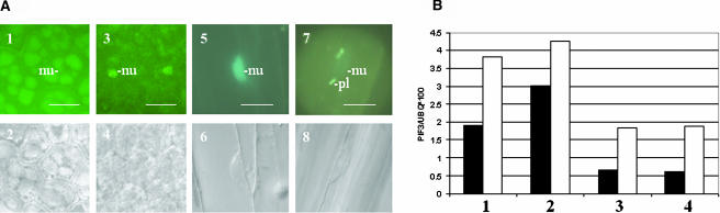
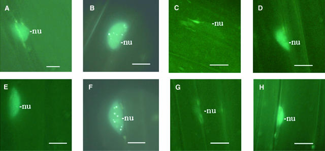
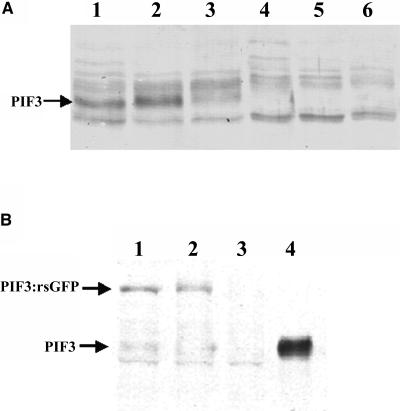
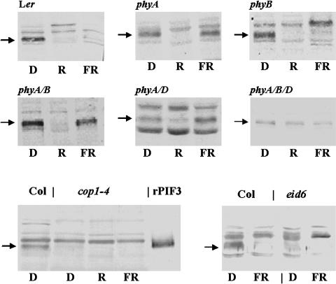
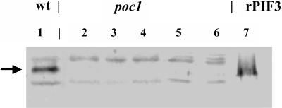
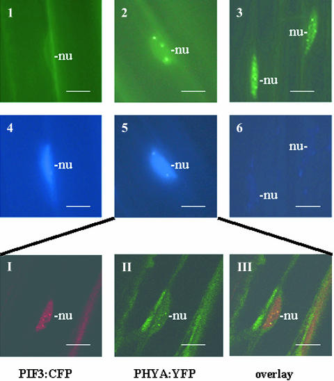
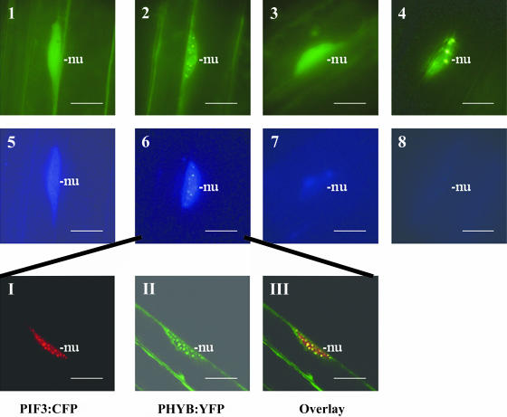
Light, in a quality- and quantity-dependent fashion, induces nuclear import of the plant photoreceptors phytochrome, promotes interaction of phytochrome A (phyA) and phyB with transcription factors including phytochrome interacting factor 3 (PIF3), and is thought to trigger a transcriptional cascade to regulate the expression of approximately 2500 genes in Arabidopsis thaliana. Here, we show that controlled degradation of the transcription factor PIF3 is a major regulatory step in light signaling. We demonstrate that accumulation of PIF3 in the nucleus in dark requires constitutive photomorphogenesis 1 (COP1), a negative regulator of photomorphogenesis, and show that red (R) and far-red light (FR) induce rapid degradation of the PIF3 protein. This process is controlled by the concerted action of the R/FR absorbing phyA, phyB, and phyD photoreceptors, and it is not affected by COP1. Rapid light-induced degradation of PIF3 indicates that interaction of PIF3 with these phytochrome species is transient. In addition, we provide evidence that the poc1 mutant, a postulated PIF3 overexpressor that displays hypersensitivity to R but not to FR, lacks detectable amounts of the PIF3 protein. Thus, we propose that PIF3 acts transiently, and its major function is to mediate phytochrome-induced signaling during the developmental switch from skotomorphogenesis to photomorphogenesis and/or dark to light transitions.


Figures

Figure 1.
Figure 1.
Overexpression and Detection of PIF3 and PIF3:rsGFP Protein in Transgenic Plants. (A) Transgenic seedlings overexpressing PIF3 or PIF3:rsGFP fusion protein are hyposensitive to cR. Transgenic Arabidopsis seedlings overexpressing PIF3 (1) or PIF3:rsGFP (2) and wild-type (ecotype Wassilewskija) (3) seedlings were grown for 4 d under 20 μmol/m2/s R, and inhibition of hypocotyl elongation was determined. Hypocotyl length as percentage of the corresponding dark control is shown. Each value represents an average of three independent experiments; error bars indicate the standard error of the means. (B) The PIF3 antibody recognizes PIF3 in total plant cell extracts. The polyclonal antiserum raised against full-length recombinant PIF3 (rPIF3) cross-reacts with other proteins but recognizes the PIF3 in total plant protein extracts. Plant extracts were prepared from 6-d-old etiolated seedlings, and 2.0 ng rPIF3 and 0 μg plant protein extract (lane 1), 0 μg rPIF3 and 20 μg plant protein extract (lane 2), and 3.0 ng rPIF3 and 20 μg plant protein extract (lane 3) were subjected to protein gel blot hybridization after SDS-PAGE. The arrow indicates the position of rPIF3 and the endogenous plant PIF3 protein. (C) Transgenic plants overexpress the PIF3 or PIF3:rsGFP fusion protein. Total protein extracts were prepared from etiolated wild-type (lane 3) and transgenic seedlings overexpressing PIF3 (lane 1) or PIF3:rsGFP fusion protein (lane 2). Accumulation of PIF3 or PIF3:rsGFP was determined by protein gel blot hybridization using the polyclonal antiserum described in Figure 1B. All lanes contain equal amounts of protein (20 μg). The arrow indicates the position of PIF3; the asterisk indicates the position of the PIF3:rsGFP fusion protein.
Figure 2.
Figure 2.
Developmentally Regulated Expression of PIF3 Is Light Independent. (A) Expression of 35S:PIF3:rsGFP is regulated developmentally. 35S:PIF3:rsGFP-expressing transgenic seedlings were germinated and grown in dark (1 to 6) or in 7 μmol/m2/s white light (7 and 8). Subcellular distribution of the PIF3:GFP fusion protein was monitored by fluorescence microscopy (1, 3, 5, and 7). Bright-field images of the cells analyzed are also shown (2, 4, 6, and 8). Accumulation level of PIF3:rsGFP in seeds 2 d after imbibition (1 and 2) and in seedlings grown 2 d in dark (3 and 4, cotyledon), for 4 d in dark (5 and 6, hypocotyl), or 4 d in light (7 and 8, hypocotyl) after germination was induced. Scale bars = 10 μm. Positions of nuclei (nu) and plastids (pl) are indicated. (B) Expression of PIF3 is not regulated by light at the level of transcription. Accumulation of PIF3 and PIF3:rsGFP mRNA was determined by RNase protection experiments. Total RNA was extracted from seeds 1 d after imbibition (1), from 2-d-old (2) and 4-d-old seedlings (3) grown in dark, or from 4-d-old etiolated seedlings irradiated with 4 h of red light before harvesting (4). Closed bars represent PIF3, and open bars represent PIF3:rsGFP steady state mRNA levels normalized to the corresponding ubiquitin (UBQ) signals.
Figure 3.
Figure 3.
Accumulation and Cellular Distribution of PIF3:CFP Is Affected by R and FR Light Treatment. Epifluorescence images of hypocotyl cells expressing PIF3:CFP in 6-d-old dark-grown wild-type seedlings ([A] and [E]), irradiated with 2 min FR (B) or R (F) and returned afterwards to darkness for 30 min ([C] and [G]) and for 6 h ([D] and [H]). Scale bars = 10 μm. Positions of nuclei (nu) are indicated.
Figure 4.
Figure 4.
Degradation of Both PIF3 and PIF3:rsGFP Is Induced by R Treatment. Total protein extracts were prepared and the accumulation level of PIF3 was determined by protein gel blot hybridization using the antiserum raised against PIF3. (A) Wild-type seedlings were grown for 6 d in dark (lane 1) and irradiated with R for 2 min (lane 2), 10 min (lane 3), 30 min (lane 4), 45 min (lane 5), and 6 h (lane 6). Each lane contains equal amounts of protein (30 μg). Position of the PIF3 is indicated by an arrow. (B) Alternatively, transgenic seedlings expressing the PIF3:rsGFP were grown for 6 d in dark (lane 1) and were irradiated with R for 5 min (lane 2) or for 3 h (lane 3). Lane 4 contains 2.0 ng of recombinant PIF3 protein; lanes 1 to 3 contain equal amounts of plant total protein extract (20 μg). The bands representing PIF3 and PIF3:rsGFP are marked.
Figure 5.
Figure 5.
FR- and R-Induced Degradation of PIF3 Is Regulated by the Concerted Action of phyA, phyB, and phyD, whereas COP1 Is Required for the Accumulation of PIF3 in Dark. Wild-type (Landsberg erecta and Columbia), phyA-201, phyB-5, phyA-201 phyB-5, phyA-201 phyD-1, phyA-201 phyB-5 phyD-1, cop1-4, and eid6 mutant seedlings were grown for 6 d in dark and then exposed to 1 h of R or FR. Total protein extracts were prepared and PIF3 abundance was determined by protein gel blot hybridization. All lanes contain identical amounts of protein extract (20 μg). D, dark; rPIF3, recombinant PIF3. Arrows mark the positions of PIF3.
Figure 6.
Figure 6.
poc1 Mutant Seedlings Do Not Contain Detectable Amounts of PIF3. Abundance of PIF3 in total protein extracts prepared from wild-type and poc1 seedlings was determined by protein gel blot hybridization. Wassilewskija wild-type (lane 1) and poc1 seedlings were germinated and grown in dark for 6 d. The 6-d-old etiolated poc1 seedlings (lane 2) were then irradiated with R for 1 h (lane 3) and 2 h (lane 4). Alternatively, 6-d-old etiolated poc1 seedlings were grown for an additional 4 d in dark (lane 5) or irradiated for 4 d with cR (lane 6). Lanes 1 to 6 contain identical amounts of plant total protein extract (20 μg), and lane 7 contains 2 ng of recombinant PIF3 (rPIF3) protein isolated from Escherichia coli. An arrow indicates the position of PIF3 and rPIF3.
Figure 7.
Figure 7.
PIF3:CFP Is Colocalized Transiently with PHYA:YFP in Nuclei of FR Light–Irradiated Transgenic Seedlings. Transgenic lines expressing both PHYA:YFP and PIF3:CFP in phyA-211 background were used to determine cellular distribution of the fusion proteins. Epifluorescent images of PhyA:YFP (1 to 3) and PIF3-CFP (4 to 6) are shown in 6-d-old etiolated seedlings (1 and 4) irradiated with 2 min of FR (2 and 5) and 20 h of cFR (3 and 6). The insert presents confocal microscopic images of cells expressing both fusion proteins after 2 min of FR treatment. PIF3:CFP is displayed in red (I), PHYA:YFP is shown by green (II), and overlay of the two signals is indicated by yellow (III). Positions of nuclei (nu) are indicated. Scale bars = 10 μm.
Figure 8.
Figure 8.
PHYB:YFP Forms Early and Late Speckles. Early PHYB:YFP speckles are colocalized transiently with PIF3:CFP speckles in the nuclei of R-irradiated transgenic seedlings. Transgenic lines expressing both PHYB:YFP and PIF3:CFP in phyB-9 background were used to determine cellular distribution of these fusion proteins. Epifluorescent images of PHYB:YFP (1 to 4) and PIF3:CFP (5 to 8) are shown in 6-d-old etiolated seedlings (1 and 5) irradiated with 2 min (2 and 6), 1 h (3 and 7), and 16 h (4 and 8) of red light. The insert shows confocal microscopic images of cells expressing both fusion proteins after 2 min of R treatment. PIF3:CFP is displayed in red (I), PHYB:YFP is shown by green (II), and overlay of the two signals is indicated by yellow (III). Positions of nuclei (nu) are indicated. Scale bars = 10 μm.
Figure 9.
Figure 9.
PHYB:GFP Does Not Form Early Speckles in poc1 Seedlings. Epifluorescent analysis of the cellular distribution of PHYB:GFP is shown in 6-d-old etiolated wild-type (1 to 4) and poc1 (5 to 8) seedlings irradiated with 2 min (2 and 6), 1 h (3 and 7), and 6 h of R (4 and 8). Positions of nuclei (nu) and plastids (pl) are indicated. Scale bars = 10 μm.

References

    1. Aukerman, M.J., Hirschfeld, M., Wester, L., Weaver, M., Clack, T., Amasino, R.M., and Sharrock, R.A. (1997). A deletion in the PHYD gene of the Arabidopsis Wassilewskija ecotype defines a role for phytochrome D in red/far-red light sensing. Plant Cell 9, 1317–1326. - PMC - PubMed
    1. Büche, C., Poppe, C., Schäfer, E., and Kretsch, T. (2000). eid1: A new Arabidopsis mutant hypersensitive in phytochrome A-dependent high-irradiance responses. Plant Cell 12, 547–558. - PMC - PubMed
    1. Chattopadhyay, S., Ang, L.H., Puente, P., Deng, X.W., and Wei, N. (1998). Arabidopsis bZIP protein HY5 directly interacts with light-responsive promoters in mediating light control of gene expression. Plant Cell 10, 673–683. - PMC - PubMed
    1. Choi, G., Yi, H., Lee, J., Kwon, Y.K., Soh, M.S., Shin, B., Luka, Z., Hahn, T.R., and Song, P.S. (1999). Phytochrome signalling is mediated through nucleoside diphosphate kinase 2. Nature 401, 610–613. - PubMed
    1. Clack, T., Matthews, S., and Sharrock, R.A. (1994). The phytochrome apoprotein family in Arabidopsis is encoded by five genes: The sequence and expression of PHYD and PHYE. Plant Mol. Biol. 25, 413–417. - PubMed

Publication types

MeSH terms