. 2004 Jun;16(6):1536-49.
doi: 10.1105/tpc.021154. Epub 2004 May 21.

NAI1 gene encodes a basic-helix-loop-helix-type putative transcription factor that regulates the formation of an endoplasmic reticulum-derived structure, the ER body

Affiliations

NAI1 gene encodes a basic-helix-loop-helix-type putative transcription factor that regulates the formation of an endoplasmic reticulum-derived structure, the ER body

Ryo Matsushima et al. Plant Cell. 2004 Jun.

Abstract

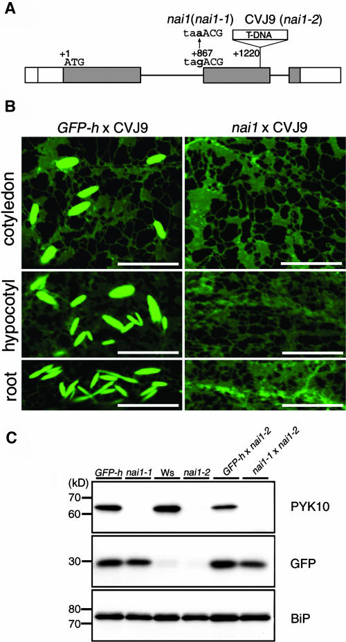
Plant cells develop various types of endoplasmic reticulum (ER)-derived structures with specific functions. ER body, an ER-derived compartment in Arabidopsis thaliana, is a spindle-shaped structure. The NAI1 gene regulates the development of ER bodies because mutation of NAI1 abolishes the formation of ER bodies. To better understand the role of NAI1, we cloned the NAI1 gene using a positional cloning strategy. The nai1-1 mutant had a single nucleotide change at an intron acceptor site of At2g22770 (NAI1 gene). Because of this mutation, aberrant splicing of NAI1 mRNA occurs in the nai1-1 mutant. NAI1 encodes a transcription factor that has a basic-helix-loop-helix (bHLH) domain. Transient expression of NAI1 induced ER bodies in the nai1-1 mutant. Two-dimensional electrophoresis and RT-PCR analyses showed that a putative lectin was depressed at both the mRNA and protein levels in nai1 mutants, as was a beta-glucosidase (PYK10). Our results provide direct evidence that a bHLH protein plays a role in the formation of ER bodies.


Figures

Figure 1.
Figure 1.
Positional Cloning of the NAI1 Gene. (A) Fine mapping of the NAI1 gene on chromosome 2. Names and positions of the molecular markers are indicated. T9I22, T30L20, and T20K9 are BAC clones. We analyzed 174 F2 progeny (348 chromosomes) having homozygous nai1 alleles. The numbers of recombinations that occurred between the NAI1 locus and the molecular markers are indicated. The NAI1 locus was mapped to a 69-kb region between two molecular markers (T9I22-4 and T30L20-4). This region contained 14 open reading frames (boxes). The nai1 mutant had a mutation in the At2g22770 gene (gray box). (B) A schematic representation of the exon and intron organization of At2g22770 and deduced protein structure. The translation start codon (ATG) is designated nucleotide +1, and the stop codon (TAA) is +1464. At2g22770 contains three exons and two introns (bars). White boxes indicate untranslated regions in exons, and gray boxes indicate translated regions in exons. Two distinct transcription start sites (arrows) were determined based on the capping structure of mRNA (see Methods). The nai1 mutant had a single base pair change (G to A) at an intron splicing acceptor site (+867). At2g22770 encodes a 320–amino acid protein that contains an acidic region (12 to 24), two Ser-rich regions (64 to 87 and 212 to 233) and a basic-helix-loop-helix domain (125 to 177). Basic, helix, and loop regions are indicted by yellow, blue, and gray boxes, respectively. (C) Alignment of amino acid sequences of the bHLH domain (125 to 177) of At2g22770 protein and other bHLH proteins, PIF3 (340 to 392, Arabidopsis), AtMYC2 (445 to 497, Arabidopsis), AN1 (468 to 520, Petunia x hybrida), DEL (436 to 488, Antirrhinum majus), PG1 (455 to 507, Phaseolus vulgaris), c-myc (351 to 406, Homo sapiens), and MAX (20 to 74, H. sapiens). These bHLH proteins are thought to function as transcription factors. The Glu residue indicated by an arrow is critical for the recognition of the E-box sequence. Identical residues are shown in black, and similar residues are shown in gray.
Figure 2.
Figure 2.
Aberrant Splicing of At2g22770 mRNA in the nai1 Mutant. (A) Analyses of At2g22770 expression in wild-type Col, GFP-h, and nai1. Actin was used as an internal control. The lane GFP-hRT− is the negative control in which reverse transcriptase was omitted from the reaction mixture. (B) Electropherogram of the amplified cDNA fragment of At2g22770 RNA resolved by capillary electrophoresis. The 35-cycle products in (A) were analyzed. The amplified cDNA fragment of nai1 had a smaller size than the amplified cDNA fragments of Col (blue) and GFP-h (black). The peak of nai1 (red) had a shoulder. The peak and shoulder positions were 9 or 16 base pairs smaller, respectively, compared with the peak positions of Col and GFP-h. (C) Effect of the single nucleotide mutation in nai1 on the translation of predicted proteins. Sequencing of the amplified cDNA fragment revealed that the G-to-A transition at the intron acceptor site resulted in aberrant splicing. Asterisks indicate the mutated nucleotide. Two distinct splicing patterns were detected in nai1 (nai1 cDNA1 and cDNA2). Boxed sequences in the genome are introns that are spliced out in the mature mRNA.
Figure 3.
Figure 3.
Allelism Test between nai1 and T-DNA Insertion Line. (A) Schematic representation of the T-DNA insertion site in CVJ9 line. (B) Fluorescent images of F1 progeny of a cross between GFP-h × CVJ9 and nai1 × CVJ9. No ER bodies were detected in the F1 seedlings of nai1 × CVJ9. Bars = 10 μm. (C) Absence of PYK10 in nai1-1, nai1-2, or F1 progeny of nai1-1 × nai1-2. Extracts prepared from 7-d-old GFP-h, nai1-1, Ws, nai1-2, and F1 progeny of GFP-h × nai1-2 and F1 progeny of nai1-1 × nai1-2 were subjected to immunoblot analyses with anti-PYK10, anti-GFP, and anti-BiP antibodies. Molecular masses are given at the left in kD.
Figure 4.
Figure 4.
Complementation of the nai1 Phenotype by Transiently Expressed At2g22770 Protein. (A) Fluorescent images of nai1-1 epidermal cells that expressed At2g22770 protein and mRFP1. Plasmid (mRFP1 + At2g22770/pBI221) from which mRFP1 and At2g22770 protein were expressed under the control of the 35S promoter was bombarded into 7-d-old nai1-1 seedlings. Images were taken with a confocal laser scanning microscope 48 to 53 h postbombardment. Spindle-shaped ER bodies were observed in the bombarded cells. Bars = 20 μm. (B) Fluorescent images of nai1-1 epidermal cells that expressed only mRFP1. Plasmid (mRFP1/pBI221) from which only mRFP1 was expressed under the control of the 35S promoter was bombarded. No ER bodies were detected in the bombarded cells. Bars = 20 μm.
Figure 5.
Figure 5.
Two-Dimensional Electrophoresis of Proteins in the P1 Fraction from GFP-h and nai1-1. Six-day-old seedlings of GFP-h and nai1-1 were homogenized and centrifuged at 1000g. The P1 fraction was separated by two-dimensional electrophoresis with denaturing isoelectric focusing (IEF) on immobilized pH gradients in the first dimension and SDS-PAGE in the second dimension. Proteins were detected by silver staining. Arrows indicate the protein spots that were specific to GFP-h. The protein spot numbers refer to the spot numbers listed in Table 1. Numbers on the x axis are pI, and numbers on the y axis are molecular mass (kD).
Figure 6.
Figure 6.
Downregulation of PYK10 and At3g16420 mRNA in nai1-1 and nai1-2 Mutants. (A) Absence of At3g16420 protein in nai1-1, nai1-2, or F1 progeny of nai1-1 × nai1-2. Immunoblot analysis was performed with anti-At3g16420 antibodies on the same extracts as Figure 3C. Molecular masses are given at the left in kD. (B) The mRNA levels of PYK10 and At3g16420 genes were monitored by RT-PCR. Total RNA from 6-d-old whole seedlings of Col, GFP-h, nai1-1, Ws, and nai1-2 was subject to reverse transcription. Actin was used as an internal control. The lanes GFP-hRT− and WsRT− are the negative controls in which reverse transcriptase was omitted from the reaction mixture.
Figure 7.
Figure 7.
Decreased β-d-Glucosidase and β-d-Fucosidase Activity in nai1-1 and nai1-2 Mutants. (A) Assay was performed using a fluorogenic substrate, 4-MU β-d-glucopyranoside, by monitoring the increase of fluorescence. β-d-Glucosidase activity of nai1-1 decreased compared with that of GFP-h and β-d-glucosidase activity of nai1-2 decreased compared with that of Ws. (B) Effect of anti-PYK10 antibodies (i) or its preimmune serum (p) on β-d-glucosidase activity. Extracts were incubated with anti-PYK10 antibodies or its preimmune serum before mixing with the substrate. The inhibitory effect of anti-PYK10 antibodies indicates that PYK10 has β-d-glucosidase activity. (C) Assay was performed using a fluorogenic substrate, 4-MU β-d-fucoside, by monitoring the increase of fluorescence. β-d-Fucosidase activity of nai1-1 decreased compared with that of GFP-h, and β-d-fucosidase activity of nai1-2 decreased compared with that of Ws. (D) Effect of anti-PYK10 antibodies (i) or its preimmune serum (p) on β-d-fucosidase activity. The inhibitory effect of anti-PYK10 antibodies indicates that PYK10 has β-d-fucosidase activity. Each bar represents the mean value of three independent experiments, and error bars show standard deviations.
Figure 8.
Figure 8.
Irregular Shapes of i-ER Bodies in nai1-1 Rosette Leaves and Induction of the NAI1 Gene by MeJA Treatment. (A) nai1-1 and GFP-h rosette leaves were treated with 50 μM MeJA or water. Epidermal cells of each rosette leaf were inspected with a confocal microscope 34 to 36 h later. Bars = 10 μm. (B) Expression of NAI1, PYK10, and At3g16420 genes in water-treated, MeJA-treated, and MeJA plus ethylene–treated leaves was monitored by RT-PCR. Actin was used as an internal control. The lane MeJART− is a negative control in which reverse transcriptase was omitted in the reaction mixture of MeJA-treated leaves.
Figure 9.
Figure 9.
Repeated Protein Structure of At3g16420 Protein Homologous to a Lectin. (A) At3g16420 protein consists of two consecutive repeated regions (gray boxes of At3g16420-N and At3g16420-C, respectively). Both regions show high homology with α-chain of jacalin, a lectin isolated from jackfruit (Artocarpus integrifolia). Myrosinase binding protein 70p (MBP70p, Brassica napus) has three jacalin-homologous regions (gray boxes of repeats 1, 2, and 3, respectively). (B) Alignment of amino acid sequences of the repeated regions of At3g16420 protein (At3g16420-N and At3g16420-C), MBP70p (70p-repeats 1, 2, and 3), and α-chain of jacalin (jacalin-α). Identical residues are shown in black, and similar residues are shown in gray.

References

    1. Abe, H., Yamaguchi-Shinozaki, K., Urao, T., Iwasaki, T., Hosokawa, D., and Shinozaki, K. (1997). Role of Arabidopsis MYC and MYB homologs in drought- and abscisic acid-regulated gene expression. Plant Cell 9, 1859–1868. - PMC - PubMed
    1. Augur, C., Benhamou, N., Darvill, A., and Albersheim, P. (1993). Purification, characterization, and cell wall localization of an α-fucosidase that inactivates a xyloglucan oligosaccarin. Plant J. 3, 415–426. - PubMed
    1. Babcock, G.D., and Esen, A. (1994). Substrate specificity of maize β-glucosidase. Plant Sci. 101, 31–39.
    1. Baumann, O., and Walz, B. (2001). Endoplasmic reticulum of animal cells and its organization into structural and functional domains. Int. Rev. Cytol. 205, 149–214. - PubMed
    1. Behnke, H.-D., and Eschlbeck, G. (1978). Dilated cisternae in Capparales—An attempt towards the characterization of a specific endoplasmic reticulum. Protoplasma 97, 351–363.

Publication types

MeSH terms