. 2004 Jun;16(6):1478-89.
doi: 10.1105/tpc.021147. Epub 2004 May 21.

Repression of AGAMOUS by BELLRINGER in floral and inflorescence meristems

Affiliations

Repression of AGAMOUS by BELLRINGER in floral and inflorescence meristems

Xiaozhong Bao et al. Plant Cell. 2004 Jun.

Abstract

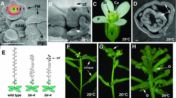
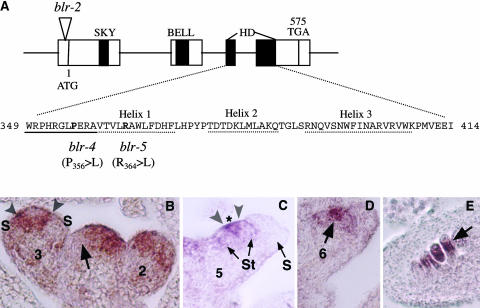
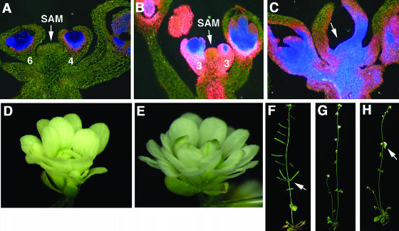
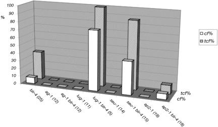
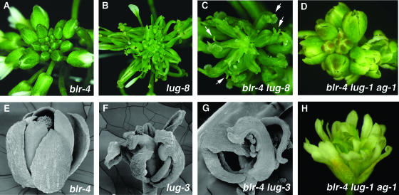
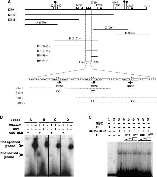
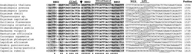
A common aspect of gene regulation in all developmental systems is the sustained repression of key regulatory genes in inappropriate spatial or temporal domains. To understand the mechanism of transcriptional repression of the floral homeotic gene AGAMOUS (AG), we identified two mutations in the BELLRINGER (BLR) gene based on a striking floral phenotype, in which homeotic transformations from sepals to carpels are found in flowers derived from old terminating shoots. Furthermore, this phenotype is drastically enhanced by growth at a high temperature and by combining blr with mutants of LEUNIG and SEUSS, two putative transcriptional corepressors of AG. We showed that the floral phenotype of blr mutants is caused by derepression of AG, suggesting that BLR functions as a transcription repressor. Because BLR encodes a BELL1-like (BELL) homeobox protein, direct binding of BLR to AG cis-regulatory elements was tested by gel-shift assays, and putative BLR binding motifs were identified. In addition, these putative BLR binding motifs were shown to be conserved in 17 of the 29 Brassicaceae species by phylogenetic footprinting. Because BELL homeobox proteins are a family of plant-specific transcription factors with 12 members in Arabidopsis thaliana, our findings will facilitate the identification of regulatory targets of other BELL proteins and help determine their biological functions. The age-dependent and high temperature-enhanced derepression of AG in blr mutants led us to propose that AG expression might be regulated by a thermal time-dependent molecular mechanism.


Figures

Figure 1.
Figure 1.
Characterization of blr Mutant Phenotypes. (A) A scanning electron microscopy photo of the tip of a wild-type shoot illustrating the SAM and FM. 1, 2, 3, and 4 indicate the four floral whorls. (B) A scanning electron microscopy photo of a blr-4 SAM exhibiting the tcf phenotype. Carpelloid flowers (cf) are indicated. A smaller SAM (arrow) is evident. (C) A wild-type flower showing the four floral organ types, sepal (S), petal (P), stamen (St), and carpel (Ca), in whorls 1, 2, 3, and 4, respectively. (D) A cross section of a typical blr-4 carpelloid flower. Arrows indicate the formation of gyneocia (g) in whorls 1 and 4. (E) A diagram summarizing the tcf and phyllotaxy defects of blr-4 mutants. Open circles indicate normal flowers, and closed circles indicate carpelloid flowers. Some blr-4 plants only exhibit phyllotaxy defects, whereas others exhibit both phyllotaxy and tcf. (F) A wild-type terminating shoot. Siliques are remnant of normal flowers. (G) A blr-4 plant exhibiting tcf (arrow). Note the abrupt transition from siliques to carpelloid flowers. (H) A blr-4 terminating shoot exhibiting more carpelloid flowers at 29°C. Carpelloid bracts subtending each flower and bearing ovules (o) are indicated by arrows. Bars in (A), (B), and (D) = 100 μm.
Figure 2.
Figure 2.
Molecular Analyses of BLR, a BELL Class Homeodomain Protein. (A) A diagram illustrating the BLR gene organization and the molecular lesions in blr-2, blr-4, and blr-5. The four exons are indicated by the four boxes connected by a line. The locations of the SKY, BELL, and homedomain (HD) are indicated by closed regions. The amino acid sequence in the homeodomain is shown. The three α helices and the N-terminal arm are underlined by dotted and solid lines, respectively. The amino acids affected by blr-4 and blr-5 are in bold. Numbers indicate the amino acid. (B) to (E) In situ hybridizations of 8-μm longitudinal sections of inflorescences using a BLR 3′ probe. This probe detected no signal in blr-2 tissues. Numbers indicate the stage of each flower. (B) In the shoot apex, an arrow indicates the exclusion of BLR mRNA from an emerging FM. In the stage 2 flower, BLR mRNA is present in the peripheral zone. In the stage 3 flower, BLR mRNA becomes excluded from the sepal primordia (S). A narrow band of BLR-expressing cells (marked by a pair of arrowheads) flanks the center of the stage 3 flower. (C) A stage 5 flower showing a narrow BLR-expressing band (marked by a pair of arrowheads) situated between stamen (St) primordia and the remaining central zone marked by an asterisk. (D) BLR is localized in the center (arrow) of a stage 6 flower. (E) BLR mRNA is detected in the chalazal (arrow) domain of developing ovules.
Figure 3.
Figure 3.
Ectopic AG Expression Mediates tcf but Not Phyllotaxy Defects of blr-4. All plants in this figure were grown at 29°C. Numbers indicate the stage of flowers. The AG:GUS reporter expression ([A] to [C]) is indicated by blue (strong) to pink (weak) staining. (A) A longitudinal section of a blr-4 shoot before exhibiting tcf. The AG:GUS reporter expression reflects normal AG mRNA expression pattern in whorls 3 and 4 of flowers at stage 4 and stage 6. (B) A blr-4 shoot at the beginning tcf phase. Ectopic AG expression is detected in all floral whorls, in SAM, and in the stem. (C) A blr-4 shoot at the later phase of tcf. SAM (arrow) becomes flattened and difficult to identify. Ectopic AG is seen throughout the FM, SAM, and the stem. (D) An ag-1 flower. (E) An ag-1 blr-4 double mutant flower resembling ag-1 flowers. (F) A blr-4 plant showing the abnormal phyllotaxy. A cluster of siliques (arrow) originates from the same position on a stem. (G) An ag-1 plant with normal phyllotaxy. (H) A blr-4 ag-1 plant showing clusters of flowers (arrow).
Figure 4.
Figure 4.
blr-4 tcf Is Enhanced by lug and seu but Not by ap2. tcf phenotype in various blr-4 double mutants. Hatched bars represent the percentage of plants with carpelloid flowers in the primary shoot. The open bars indicate the average percentage of carpelloid flowers per inflorescence shoot that exhibits a tcf phenotype. Only primary shoot is used in the analysis. Numbers in the parentheses indicate the number of plants scored. All plants were grown at 20°C except ag-1 and ag-1 blr-4 plants, which were grown at 29°C.
Figure 5.
Figure 5.
blr-4 Exhibits Synergistic Genetic Interactions with lug. All plants in this figure were grown at 20°C. (A) A blr-4 young inflorescence. At this early stage and at 20°C, most plants do not exhibit any tcf and are similar to wild-type inflorescences. (B) A lug-8 inflorescence with flowers exhibiting narrow sepals and reduced numbers of petals. (C) A blr-4 lug-8 double mutant inflorescence showing a dramatically enhanced floral phenotype. Most carpelloid organs are topped with horns (arrows). (D) A blr-4 lug-1 ag-1 triple mutant inflorescence showing no sign of tcf. (E) A blr-4 flower developed from a young inflorescence at 20°C. It resembles wild-type flowers. (F) A lug-3 flower showing partially carpelloid sepals and an absence of petals. (G) A blr-4 lug-3 double mutant flower showing carpelloid whorl 1 organs. (H) An ag-1 blr-4 lug-1 triple mutant flower resembling ag-1 flowers. However, floral organs of the triple mutant are narrower than those of ag-1.
Figure 6.
Figure 6.
BLR Directly Binds to the 3-kb AG Intron. (A) A diagram summarizing the mobility-shift assay results. The ∼3-kb AG HindIII fragment used in the KB9 reporter roughly coincides with the AG second intron. Numbers above the KB9 line indicate the nucleotide sequence with the 5′ HindIII site designated 1. AG intron fragments used in KB14 and KB11 reporters (Busch et al., 1999) are indicated. Closed circles indicate the location of previously identified LFY/WUS binding sites (Hong et al., 2003). Closed triangles indicate the location of BBS. The A, B, C, D, B1, B7, B8, and B9 fragments served as probes. The size of each fragment is indicated by a number within parentheses. Plus and minus signs indicate positive and negative mobility shifts, respectively. BBS1-3 is underlined beneath the sequence within the B9 fragment. The orientation of each BBS is indicated by an arrow. The sequence of B13 and B10 oligos are indicated by dotted lines. B13m and B10m are mutant oligos with mutations introduced in the ATTA core sequence. The asterisk marks the specific nucleotide changed in B13m and B10m. (B) A representative gel-shift using the A, B, C, and D DNA fragments as probes. After incubating the corresponding hot probe with GST or GST-BLR proteins, DNase I was added to digest naked DNA. Only fragment B showed a band protected from DNase I digestion by GST-BLR. (C) A gel-shift assay using 32P-labeled B7 DNA as a probe and cold B13 or B10 oligos as competitors. B, protein extracts that do not contain GST-BLR; C, cold competitors. The presence or absence of protein extracts or oligo competitors is indicated by plus or minus signs above each lane. Lanes 4, 5, 7, and 8 show the effect of increasing amounts of cold B13 and B10 oligos, respectively, at 0.02 pmol (lanes 4 and 7) and 2 pmol (lanes 5 and 8). mB13 (lane 6) and mB10 (lane 9) represent mutant B13 and B10 oligos as cold competitors at 2 pmol. GST alone (lane 2) and protein extracts from E. coli that did not express GST-BLR (lane 1) served as negative controls.
Figure 7.
Figure 7.
DNA Sequence Alignment in the BBS1-3 Region of the AG Intron in 17 Brassicaceae Species. Conserved nucleotides are in bold, and the asterisk marks the most conserved positions. BBS1, BBS2, and BBS3 are shaded. The CCAAT box and the conserved TTCATTtACc motif are underlined twice. Putative WUS and LFY binding sites are underlined with dotted and solid lines, respectively. The number at the end of each sequence indicates the position of the first nucleotide in BBS2 (indicated by an arrow) relative to the 5′ end of the AG intron (44 bp downstream of the 5′ HindIII site shown in Figure 6).

Similar articles

Cited by

References

    1. Aukerman, M.J., and Sakai, H. (2003). Regulation of flowering time and floral organ identity by a microRNA and its APETALA2-like target genes. Plant Cell 15, 2730–2741. - PMC - PubMed
    1. Barolo, S., and Posakony, J.W. (2002). Three habits of highly effective signaling pathways: Principles of transcriptional control by developmental cell signaling. Genes Dev. 16, 1167–1181. - PubMed
    1. Baurle, I., and Laux, T. (2003). Apical meristems: The plant's fountain of youth. Bioessays 25, 961–970. - PubMed
    1. Bellaoui, M., Pidkowich, M.S., Samach, A., Kushalappa, K., Kohalmi, S.E., Modrusan, Z., Crosby, W.L., and Haughn, G.W. (2001). The Arabidopsis BELL1 and KNOX TALE homeodomain proteins interact through a domain conserved between plants and animals. Plant Cell 13, 2455–2470. - PMC - PubMed
    1. Blazquez, M.A., Soowal, L.N., Lee, I., and Weigel, D. (1997). LEAFY expression and flower initiation in Arabidopsis. Development 124, 3835–3844. - PubMed

Publication types

MeSH terms