. 2004 Jun 1;101(22):8342-7.
doi: 10.1073/pnas.0402254101. Epub 2004 May 21.

Evidence for assembly of prions with left-handed beta-helices into trimers

Affiliations

Evidence for assembly of prions with left-handed beta-helices into trimers

Cédric Govaerts et al. Proc Natl Acad Sci U S A. .

Abstract

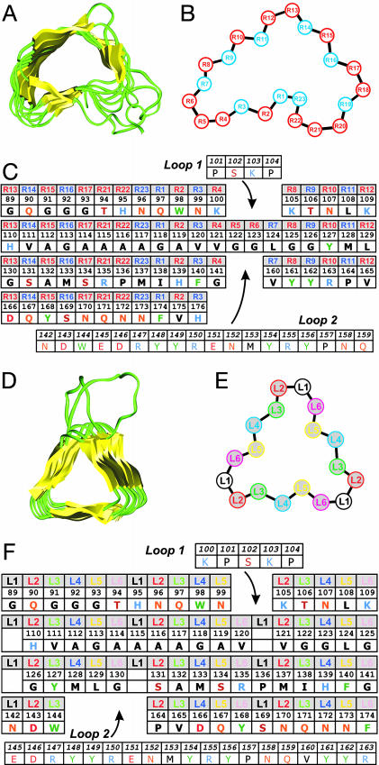
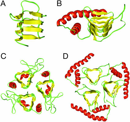
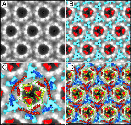
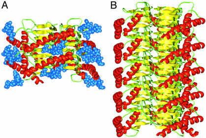
Studies using low-resolution fiber diffraction, electron microscopy, and atomic force microscopy on various amyloid fibrils indicate that the misfolded conformers must be modular, compact, and adopt a cross-beta structure. In an earlier study, we used electron crystallography to delineate molecular models of the N-terminally truncated, disease-causing isoform (PrP(Sc)) of the prion protein, designated PrP 27-30, which polymerizes into amyloid fibrils, but we were unable to choose between a trimeric or hexameric arrangement of right- or left-handed beta-helical models. From a study of 119 all-beta folds observed in globular proteins, we have now determined that, if PrP(Sc) follows a known protein fold, it adopts either a beta-sandwich or parallel beta-helical architecture. With increasing evidence arguing for a parallel beta-sheet organization in amyloids, we contend that the sequence of PrP is compatible with a parallel left-handed beta-helical fold. Left-handed beta-helices readily form trimers, providing a natural template for a trimeric model of PrP(Sc). This trimeric model accommodates the PrP sequence from residues 89-175 in a beta-helical conformation with the C terminus (residues 176-227), retaining the disulfide-linked alpha-helical conformation observed in the normal cellular isoform. In addition, the proposed model matches the structural constraints of the PrP 27-30 crystals, positioning residues 141-176 and the N-linked sugars appropriately. Our parallel left-handed beta-helical model provides a coherent framework that is consistent with many structural, biochemical, immunological, and propagation features of prions. Moreover, the parallel left-handed beta-helical model for PrP(Sc) may provide important clues to the structure of filaments found in some other neurodegenerative diseases.


Figures

Fig. 1.
Fig. 1.
Threading the PrP sequence onto β-helical architectures. (A) The structure of pectin lyase from Aspergillus niger (PDB ID code 1QCX) is shown as an example of a parallel right-handed β-helix. (B) Schematic diagram of the right-handed β-helical repeat. Positions in blue face the interior. Regions R4–R8 and R18–R21 are not conserved and may vary in length and conformation. (C) Threading of PrP residues 89–176 onto a right-handed β-helical fold. Each row represents a helical rung, the positions are labeled as in B. The template used here does not include residues at positions R18, R19, and R20, like the example shown in A. Inserted loops are indicated. Amino acids are indicated by their one-letter codes. (D) The structure of UDP N-acetylglucosamine O-acyltransferase from Escherichia coli (PDB ID code 1LXA) is shown as an example of a parallel left-handed β-helix. (E) Schematic diagram of the left-handed β-helical repeat. Each rung is made of six different positions repeated three times. Positions L3 and L5 face the interior. (F) Threading of PrP residues 89–175 onto a left-handed β-helical fold. Each row represents a helical rung; the positions are labeled as in E. Inserted loops are indicated. The absence of residues at L1 indicates short turns.
Fig. 2.
Fig. 2.
Modeling PrP residues 89–174 onto a left-handed β-helical fold. (A) The β-helical model of the N-terminal part of PrP 27–30 corresponding to the threading of Fig. 1F. (B) Model of the monomer of PrP 27–30. The α-helical region (residues 177–227) as determined by NMR spectroscopy (PDB ID code 1QM0) was linked to the β-helical model shown in A.(C) The crystal structure of the trimeric carbonic anhydrase from Methanosarcina thermophila (PDB ID code 1THJ). (D) Trimeric model of PrP 27–30 built by superimposing three monomeric models onto the coordinates of the Cα's of the 1THJ structure.
Fig. 3.
Fig. 3.
Projection map of PrP 27–30 and statistically significant differences from PrPSc106. (A) Projection map of PrP 27–30 obtained by processing and averaging three independent 2D crystals of PrP 27–30. (B) Statistically significant differences between PrP 27–30 and PrPSc106 overlaid onto the projection map of PrP 27–30. The differences attributed to the internal deletion of PrPSc106 (residues 141–176) are shown in red; the differences in glycosylation between PrP 27–30 and PrPSc106 are shown in blue. (C) Superimposition of the trimeric left-handed model onto the EM maps. The trimeric left-handed β-helical model of PrP 27–30 is superimposed on a 1:1 scale (bar = 50 Å) with the electron crystallographic maps of PrP 27–30. For the sugars linked to N180 and N196 shown as blue space-filling spheres, only the conserved core region (two N-acetylglucosamine and three mannose molecules) is depicted. Sensible side chain dihedral angles for the asparagines and oligosaccharides were selected to optimize the fit with the EM maps. (D) The scaled trimeric model was copied onto the neighboring units of the crystals (bar = 50 Å) to show the crystallographic packing suggested by the model.
Fig. 4.
Fig. 4.
Fibrillization of the trimeric left-handed β-helical discs. (A) Two discs of PrP 27–30 can assemble through polar backbone interactions between the lower β-helical rung of the top disk and the upper rung of the bottom disk. This assembly provides enough room for the α-helices to stack and the N-linked sugars to extend away from the center of the structure. (B) A model for the PrP 27–30 fiber was constructed by assembling five trimeric discs. For clarity, the sugars were omitted.

References

    1. Prusiner, S. B., Scott, M. R., DeArmond, S. J. & Cohen, F. E. (1998) Cell 93, 337-348. - PubMed
    1. Kelly, J. W. (1998) Curr. Opin. Struct. Biol. 8, 101-106. - PubMed
    1. Dobson, C. M. (2001) Philos. Trans. R. Soc. London B 356, 133-145. - PMC - PubMed
    1. Sunde, M., Serpell, L. C., Bartlam, M., Fraser, P. E., Pepys, M. B. & Blake, C. C. (1997) J. Mol. Biol. 273, 729-739. - PubMed
    1. Sunde, M. & Blake, C. C. F. (1998) Q. Rev. Biophys. 31, 1-39. - PubMed

Publication types