. 2004 May 21;304(5674):1170-3.
doi: 10.1126/science.1095211.

Definition of a bacterial type IV secretion pathway for a DNA substrate

Affiliations

Definition of a bacterial type IV secretion pathway for a DNA substrate

Eric Cascales et al. Science. .

Abstract

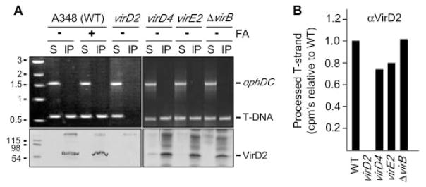
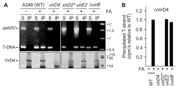
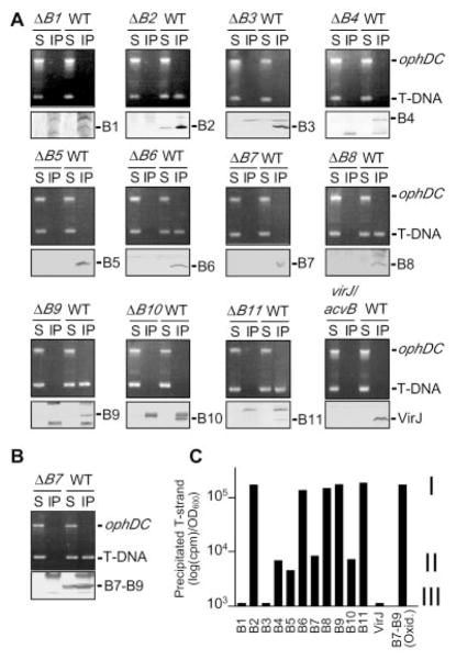
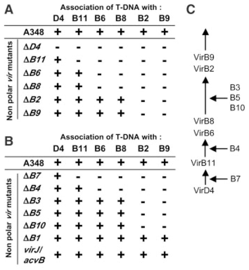
Bacteria use conjugation systems, a subfamily of the type IV secretion systems, to transfer DNA to recipient cells. Despite 50 years of research, the architecture and mechanism of action of the channel mediating DNA transfer across the bacterial cell envelope remains obscure. By use of a sensitive, quantifiable assay termed transfer DNA immunoprecipitation (TrIP), we identify contacts between a DNA substrate (T-DNA) and 6 of 12 components of the VirB/D4 conjugation system of the phytopathogen Agrobacterium tumefaciens. Our results define the translocation pathway for a DNA substrate through a bacterial conjugation machine, specifying the contributions of each subunit of the secretory apparatus to substrate passage.


Figures

Fig. 1
Fig. 1. Isolation of the VirD2–T-strand transfer intermediate
(A) Antibodies to VirD2 precipitated VirD2 detected by SDS–polyacrylamide gel electrophoresis and immunostaining (lower panels) and the T-DNA substrate (T-DNA) but not the pTi control (ophDC) fragment detected by PCR amplification and gel electrophoresis (upper panels). Strain A348 (wild-type, WT) and the isogenic mutants (table S1) were untreated (−) or treated (+) with formaldehyde (FA) before lysis. S, supernatant after immunoprecipitation; IP, immunoprecipitate. Upper bands in the immunoblots are immunoreactive immunoglobulin G (IgG) heavy chain. Molecular mass markers (left lane) are in kilobases (upper) or kilodaltons (lower). (B) T-strand levels in material precipitated with antibodies to VirD2 (αVirD2) from the strains indicated, as determined by quantitative TrIP. Data are presented as counts per minute (CPM) of incorporated radionucleotide during one cycle in the logarithmic phase of PCR amplification. Values for the mutant strains are presented as a fraction of WT, normalized to 1.0.
Fig. 2
Fig. 2. VirD4 binds the VirD2–T-strand
(A) Antibodies to VirD4 coprecipitated VirD4 (lower panels) and the T-DNA substrate (upper panels) from extracts of formaldehyde-treated (+) strain A348 (WT) and mutants defective for synthesis of the T4SS or the VirE2 SSB. virD2* is a virD2 mutant expressing virD4 from an IncP replicon. Lower bands in the immunoblots are IgG heavy chain. (B) T-strand levels in material precipitated with antibodies to VirD4 from the strains indicated, as determined by quantitative TrIP. Data are presented as CPM of incorporated radionucleotide during one cycle in the logarithmic phase of PCR amplification. Values for the mutant strains are presented as a fraction of WT, normalized to 1.0.
Fig. 3
Fig. 3. VirB/D4 T4SS subunits that associate with the T-DNA transfer intermediate
(A) Antibodies to VirB2, VirB6, VirB8, VirB9, and VirB11 coprecipitated the cognate protein (lower panels) and the T-DNA substrate (upper panels) from extracts of formaldehyde-treated A348 (WT) but not from extracts of the corresponding virB null mutant (ΔBx). ophDC, pTi control fragment; S, supernatant; IP, immunoprecipitate. (B) Under oxidizing conditions, antibodies to VirB7 coprecipitated a disulfide–cross-linked VirB7-VirB9 dimer (lower panel; blot development with antibodies to VirB7) and the T-DNA substrate (upper panel). (C) T-strand levels in material precipitated from the WT strain with antibodies to the Vir protein listed at the bottom, as determined by quantitative TrIP. Data are presented as CPM of incorporated radionucleotide during one cycle of PCR amplification on a log scale. Vir proteins were classified according to the relative level of coprecipitated T-strand: class I, abundant; class II, low; class III, undetectable.
Fig. 4
Fig. 4. Nonpolar virB and virD4 null mutations block T-strand translocation
(A) TrIP summary for mutants lacking one of the class I channel subunits; (+) denotes a T-strand interaction with the protein listed at the top; (−), no interaction. (B) TrIP summary for mutants lacking one of the class II or class III subunits. (C) A postulated contact pathway for the T-strand during translocation through the A. tumefaciens VirB/D4 T4SS. Proteins listed at right are required for passage of the T-strand at the step indicated.

Comment in

References

    1. Cascales E, Christie PJ. Nature Rev Microbiol. 2003;1:137. - PMC - PubMed
    1. An updated list of type IV secretion systems is available at http://mmg.uth.tmc.edu/webpages/faculty/supplements/pchristie/T4SS-updated.pdf.

    1. Hacker J, Hentschel U, Dobrindt U. Science. 2003;301:790. - PubMed
    1. Lawley TD, Klimke WA, Gubbins MJ, Frost LS. FEMS Microbiol Lett. 2003;224:1. - PubMed
    1. Gelvin SB. Microbiol Mol Biol Rev. 2003;67:16. - PMC - PubMed

Publication types