. 2004 May 27;42(4):567-80.
doi: 10.1016/s0896-6273(04)00255-7.

New synaptic bouton formation is disrupted by misregulation of microtubule stability in aPKC mutants

Affiliations

New synaptic bouton formation is disrupted by misregulation of microtubule stability in aPKC mutants

Catalina Ruiz-Canada et al. Neuron. .

Abstract

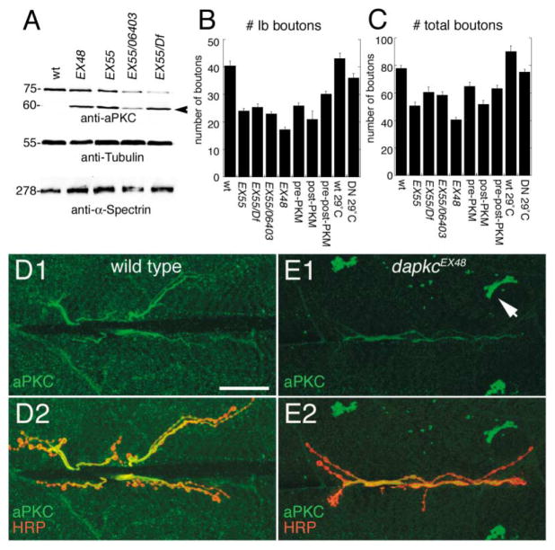
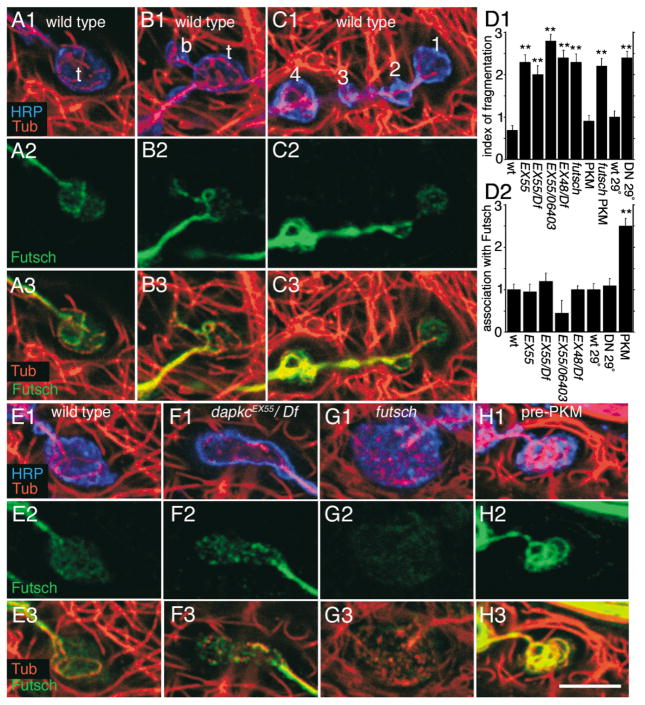
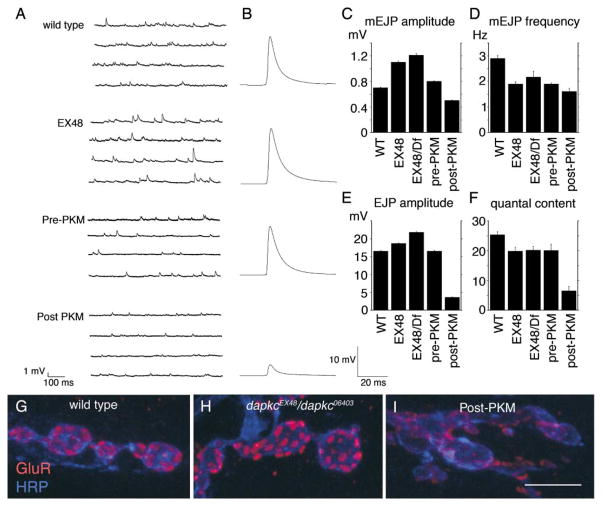
The Baz/Par-3-Par-6-aPKC complex is an evolutionarily conserved cassette critical for the development of polarity in epithelial cells, neuroblasts, and oocytes. aPKC is also implicated in long-term synaptic plasticity in mammals and the persistence of memory in flies, suggesting a synaptic function for this cassette. Here we show that at Drosophila glutamatergic synapses, aPKC controls the formation and structure of synapses by regulating microtubule (MT) dynamics. At the presynapse, aPKC regulates the stability of MTs by promoting the association of the MAP1Brelated protein Futsch to MTs. At the postsynapse, aPKC regulates the synaptic cytoskeleton by controlling the extent of Actin-rich and MT-rich areas. In addition, we show that Baz and Par-6 are also expressed at synapses and that their synaptic localization depends on aPKC activity. Our findings establish a novel role for this complex during synapse development and provide a cellular context for understanding the role of aPKC in synaptic plasticity and memory.


Figures

Figure 1
Figure 1. aPKC Is Expressed at the NMJ
(A) View of an NMJ branch labeled with (A1) anti-aPKC and (A2) anti-aPKC and anti-HRP. Arrows and arrowheads delimit a segment of the branch to show that aPKC is punctate at distal boutons. (B) NMJs at muscles 6 and 7 of a third instar larva triple labeled with anti-aPKC, Futsch, and HRP. (C) High-magnification view of the distal end of an NMJ branch in a preparation triple labeled with antibodies against aPKC, HRP, and tyr-Tub. (C1) The distribution of aPKC is shown in relation to the presynaptic compartment labeled with anti-HRP. Note that aPKC is absent from the postsynaptic peribouton area (arrow). (C2) The distribution of aPKC in relation to synaptic MTs is shown. Note that few postsynaptic MTs extend into the aPKC-free peribouton area (arrow). (D) High-magnification view of a different NMJ branch triply stained with antibodies against Spectrin, tyr-Tub, and HRP. Note that Spectrin is enriched in the postsynaptic peribouton area (arrow). Scale bar equals 16 μm in (A), 30 μm in (B), and 7.5 μm in (C) and (D).
Figure 2
Figure 2. Mutations in dapkc Lead to Abnormal aPKC Distribution and a Decrease in Synaptic Bouton Number
(A) Western blot of body wall muscle extracts in wild-type and dapkc mutants probed with anti-aPKC, anti-tubulin, and anti-α-Spectrin. Arrowhead points to an extra immunoreactive band in dapkc mutants. Numbers at left are molecular weights in kDa. (B and C) Number of type Ib (B) and total number (C) of boutons in the third instar stage (muscles 6 and 7; abdominal segment 3) in the following genotypes: wild-type (wt; n = 20), dapkcEX55 (EX55; n = 20), dapkcEX55/Df (EX55/Df; n = 20), dapkcEX55/dapkc06403 (EX55/ 06403; n = 28), dapkcEX48 (EX48; n = 22), pre-PKM (n = 23), post-PKM (n = 18), pre-post-PKM (n = 23), wild-type raised at 29°C (wt 29°C; n = 20), and PKM8-DN raised at 29°C (DN 29°C, n = 21). (D and E) NMJ at muscles 6 and 7 of (D) wild-type and (E) dapkcEX48 labeled with anti-HRP and anti-aPKC. Note that in dapkc mutants, immunoreactivity at the NMJ is reduced, and large aPKC immunoreactive aggregates are observed in the muscle cells (arrow). Scale bar equals 25 μm.
Figure 3
Figure 3. MT Cytoskeleton Organization at Distal Synaptic Boutons of Wild-Type and dapkc Mutants
Single confocal slices through wild-type distal boutons triple labeled with anti-tyr-Tubulin, anti-HRP, and anti-Futsch. (A) Wild-type terminal bouton (t). Note that Futsch is only partially colocalized with MTs at this terminal bouton. (B) Wild-type terminal bouton (t) in the process of budding (b = bud). Note that MTs extend into the bud, and Futsch colocalizes with these but not with other MTs. (C) Wild-type NMJ branch in which boutons are labeled 1 to 4 (distal to proximal). Splayed MTs are observed at both the terminal bouton (position 1) as well as at a more proximal bouton (position 4). However, Futsch is in complete colocalization with MTs only at proximal boutons (positions 2–4). (E–H) Terminal boutons from (E) wild-type, (F) dapkcEX55/Df, (G) futschk68, and (H) larvae overexpressing PKM presynaptically (pre-PKM). Note that at terminal boutons from dapkcEX55/Df, MTs are fragmented, and there is little colocalization with Futsch. This phenotype resembles the phenotype in futschK68 mutants. Also note that at terminal boutons of pre-PKM larvae, unlike wild-type, MTs are almost completely associated with Futsch. Scale bar equals 5 μm. (D1) Index of MT fragmentation, and (D2) degree of MT association with Futsch in the following genotypes: wild-type (n = 144), dapkcEX55 (n = 32), dapkcEX55/dapkc06403 (n = 22), dapkcEX55/Df (n = 28), dapkcEX48/Df (n = 30), futsch (32); pre-PKM (n = 24). futsch; pre-PKM (46); wt 29°C (n = 18); Pre-DNPKM 29°C (n = 34). n = number of boutons analyzed. **p < 0.0001.
Figure 4
Figure 4. Coimmunoprecipitation of Tubulin and Futsch by aPKC and Abnormal aPKC Localization in futsch Mutants
(A) Immunoprecipitation of adult fly head extracts with anti-aPKC in wild-type and futschk68 mutants. Proteins were resolved in a 4%–15% gradient SDS-PAGE gel prior to immunoblotting. The blot was sequentially probed with anti-aPKC, anti-Futsch, anti-Tubulin, anti-Elav, and anti-Appl. Numbers at the right are molecular weights in kDa. Input lanes correspond to 1/10 of the total extract. (B and C) NMJ branches in (B) wild-type and (C) futschk68 mutants stained with anti-aPKC, Futsch, and HRP as indicated in individual panels. Note that aPKC localization at the presynaptic MT bundle is disrupted in futsch mutants. Scale bar equals 12 μm.
Figure 5
Figure 5. Organization of Postsynaptic MTs and Spectrin at Terminal Boutons of Wild-Type and dapkc Mutants
Confocal images of NMJ branches in (A and D) wild-type, (B and E) dapkcEX55/Df, (C and F) larvae overexpressing PKM postsynaptically (post-PKM), stained with (A–C) anti-Spectrin, and (D–F) triple labeled with antibodies against tyr-Tub, HRP, and Spectrin. Arrows in (D1), (E1), and (F1) point to the peribouton area. Note that in dapkcEX55/Df, MTs are in closer proximity to the presynaptic bouton than in wild-type and that this is accompanied by a decrease in the thickness of the Spectrin-rich area (E2). Also note that MTs are significantly sparser in post-PKM larvae and that this is accompanied by an expansion of the Spectrin-rich peribouton area (F2). Scale bar equals 16 μm for (A)–(C) and 6 μm for all others.
Figure 6
Figure 6. Electrophysiological Analysis and GluR Distribution in dapkc Mutants
(A) Representative traces of mEJPs in wild-type, dapkcEx48 mutants (EX48), pre-PKM, and post-PKM. (B) Evoked EJPs in the same genotypes as in (A). Each trace is the average of 300 EJPs. (C–F) Histograms showing (C) mEJP amplitude, (D) mEJP frequency, (E) evoked EJP amplitude, and (F) quantal content in wild-type, dapkc mutants, and PKM overexpressors. Quantitative analysis was based on 5 larvae, one cell each (300 EJPs; 800 mEJPs) for each genotype. Bars represent mean ± SEM. (G–I) Distribution of GluRs in (G) wild-type, (H) dapkcEX48/dapkc06403, and (I) post-PKM NMJs. Preparations were labeled with anti-GluRIIA and anti-HRP. Scale bar is 6 μm.
Figure 7
Figure 7. Baz and Par-6 Are Localized at the NMJ in Partial Colocalization with aPKC
(A and B) Confocal images of wild-type NMJ branches triply labeled with anti-aPKC, anti-Par-6, and anti-HRP. (B3) is a high-magnification view of the box shown in (B2). (C) Confocal image of wild-type NMJs double labeled with anti-Baz and anti-Par-6. Scale bar equals 17 μm and 9 μm in (B3).
Figure 8
Figure 8. Baz and Par-6 Exist in an Endogenous Complex with aPKC, They Genetically Interact with aPKC during NMJ Expansion, and Their Localization Is Affected by Changes in aPKC Activity
(A) Coimmunoprecipitation of aPKC and Baz by anti-Par-6 antibodies in body wall muscle extracts. Blots were sequentially probed with anti-aPKC, anti-Par-6, anti-Baz, and anti-APPL. Input lanes correspond to 1/10 of the total extract. Control lane corresponds to extracts processed as the IP lanes but in which the Par-6 immunoprecipitating antibody was omitted. (B and C) NMJs in stained with anti-Baz and anti-HRP in (B) wild-type and (C) dapkcEX48. Note that both the levels and distribution of Baz is altered in dapkc mutants. (D) Total number of boutons at NMJs of dapkcEX55 (n = 21), baz4/+ (n = 23), par-6/+ (n = 22), baz4/+; dapkcEX55 (n = 20), par-6/+; dapkcEX55 (n= 20). (E–J) Single confocal slices of synaptic boutons double stained with (E, F, H, and I) anti-Baz and anti-HRP and (G and J) anti-Par-6 and anti-HRP in (E and G) wild-type, (H and J) post-PKM, (F) pre-DN-PKM (pre-DN), and (I) post-DN-PKM (post-DN). Note that post-PKM results in an expansion of peribouton Baz, while expression of the post-DN results in decrease of Baz accumulation in the presynaptic boutons (pre-DN) or peribouton area (post-DN). Scale bar equals 4 μm for all panels except (B) and (C), which is 13 μm.

Similar articles

Cited by

References

    1. Banan A, Zhang L, Fields JZ, Farhadi A, Talmage DA, Keshavarzian A. PKC-zeta prevents oxidant-induced iNOS upregulation and protects the microtubules and gut barrier integrity. Am J Physiol Gastrointest Liver Physiol. 2002;283:G909–G922. - PubMed
    1. Brand AH, Perrimon N. Targeted gene expression as a means of altering cell fates and generating dominant phenotypes. Development. 1993;118:401–415. - PubMed
    1. Budnik V, Koh YH, Guan B, Hartmann B, Hough C, Woods D, Gorczyca M. Regulation of synapse structure and function by the Drosophila tumor suppressor gene dlg. Neuron. 1996;17:627–640. - PMC - PubMed
    1. Cox DN, Seyfried SA, Jan LY, Jan YN. Bazooka and atypical protein kinase C are required to regulate oocyte differentiation in the Drosophila ovary. Proc Natl Acad Sci USA. 2001;98:14475–14480. - PMC - PubMed
    1. Dent EW, Callaway JL, Szebenyi G, Baas PW, Kalil K. Reorganization and movement of microtubules in axonal growth cones and developing interstitial branches. J Neurosci. 1999;19:8894–8908. - PMC - PubMed

Publication types

MeSH terms

LinkOut - more resources