. 2004 May 24;165(4):505-15.
doi: 10.1083/jcb.200307164.

MuSK is required for anchoring acetylcholinesterase at the neuromuscular junction

Affiliations

MuSK is required for anchoring acetylcholinesterase at the neuromuscular junction

Annie Cartaud et al. J Cell Biol. .

Abstract

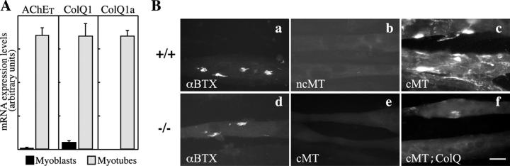
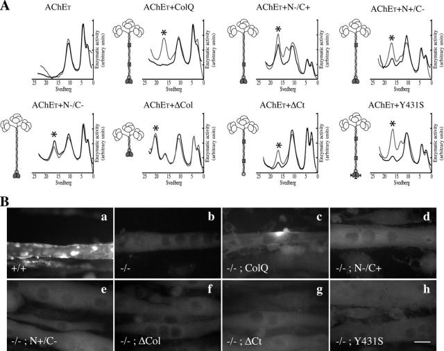
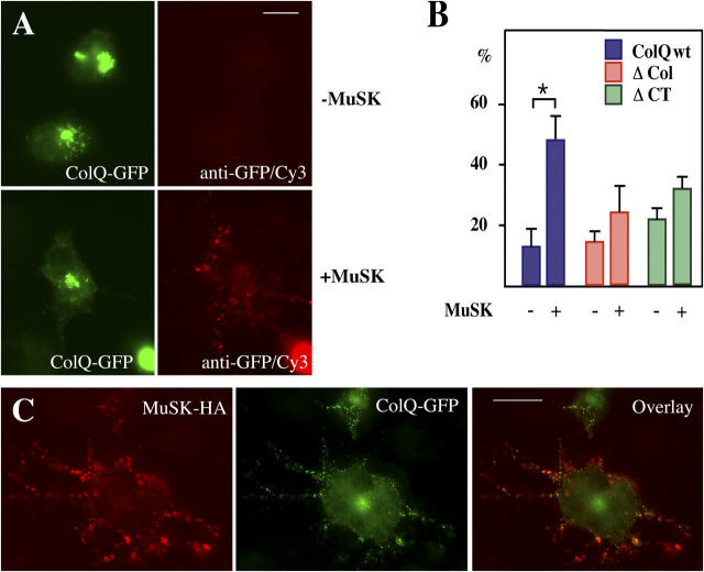
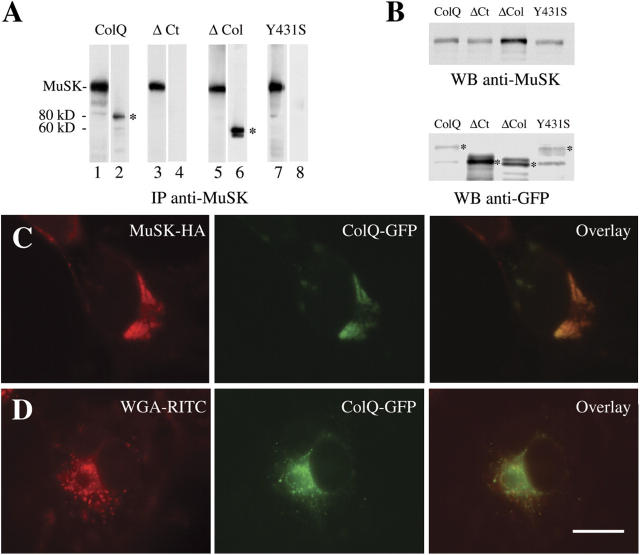
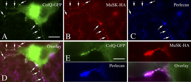
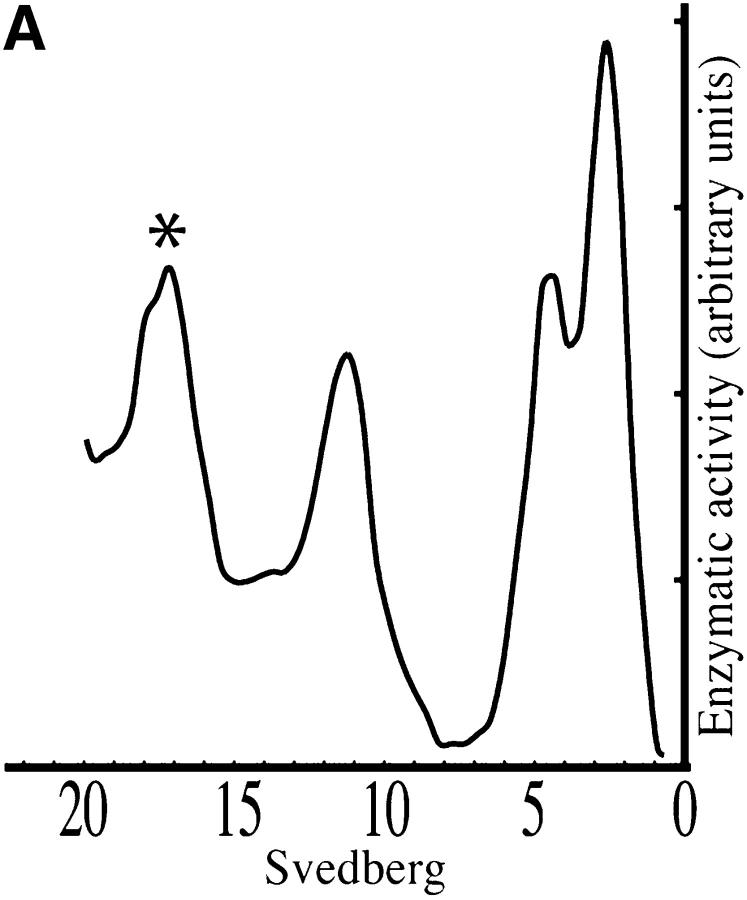
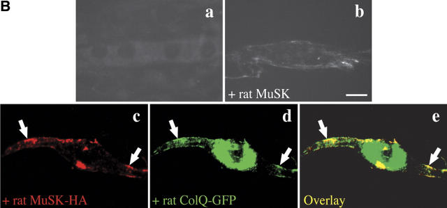
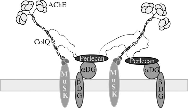
At the neuromuscular junction, acetylcholinesterase (AChE) is mainly present as asymmetric forms in which tetramers of catalytic subunits are associated to a specific collagen, collagen Q (ColQ). The accumulation of the enzyme in the synaptic basal lamina strictly relies on ColQ. This has been shown to be mediated by interaction between ColQ and perlecan, which itself binds dystroglycan. Here, using transfected mutants of ColQ in a ColQ-deficient muscle cell line or COS-7 cells, we report that ColQ clusterizes through a more complex mechanism. This process requires two heparin-binding sites contained in the collagen domain as well as the COOH terminus of ColQ. Cross-linking and immunoprecipitation experiments in Torpedo postsynaptic membranes together with transfection experiments with muscle-specific kinase (MuSK) constructs in MuSK-deficient myotubes or COS-7 cells provide the first evidence that ColQ binds MuSK. Together, our data suggest that a ternary complex containing ColQ, perlecan, and MuSK is required for AChE clustering and support the notion that MuSK dictates AChE synaptic localization at the neuromuscular junction.


Figures

Figure 1.
Figure 1.
Expression of AChE and ColQ in mouse wt and ColQ-deficient myogenic cells in culture. (A) AChET, ColQ1, and ColQ1a mRNAs levels were quantified by real-time RT-PCR (±SEM) in wt myoblasts and MTs. AChET mRNAs levels were increased by 130-fold average and ColQ1 mRNAs levels by 20-fold average after differentiation into MTs. ColQ1a transcripts were only detectable in MTs. Note that the bars do not allow direct comparison between the different transcripts. (B) Cultures were stained with either α-bungarotoxin (a and d, αBTX) or the A63 polyclonal rat anti-AChE antibody (b, c, e, and f). Wt and ColQ-deficient MTs formed AChR clusters (a and d). AChE clusters did not form in noncontracting MTs (b, ncMT) and were only revealed in contracting MTs (c, cMT). No AChE clusters were detected in ColQ-deficient contracting MTs (e). Transfection of ColQ-deficient MTs with rat ColQ1a restores the ability of these cells to form AChE clusters in contracting MTs (f). Bar, 20 μm.
Figure 2.
Figure 2.
Distinct sites of ColQ are necessary for the formation of AChE clusters. (A) Analysis of AChE molecular forms secreted from the ColQ-deficient myogenic cells transfected with AChET or cotransfected with AChET and wt ColQ or ColQ mutants. Sedimentation profiles of the molecular forms are shown in black lines. Asterisk indicates the position of A12 forms in the sedimentation profiles. Co-transfected cells were also treated for 24 h by heparin. Sedimentation profiles of the molecular forms secreted after heparin treatment are shown in gray lines. A schematic representation of AChE A forms and ColQ mutant constructs used in this work are shown on the left side of the gradients. Catalytic subunits (ovoids) are associated to a triple helix of ColQ. This last protein contains a collagen domain (186–285 aa) flanked by a NH2-terminal peptide (1–186 aa) and a COOH-terminal peptide (285–450 aa). The two HBS (N+ and C+) contained in the collagen domain are indicated as gray boxes. In mutant construct N/C+, RK (121–122 aa) is mutated to DP. In mutant construct N+/C, KR (226–227 aa) is mutated to DP. In construct N/C, both sites are mutated. In mutant ΔCol, amino acids 105–276 were deleted. In mutant ΔCt, amino acids 362–450 were deleted. The rat Y431S construct reproduces the missense mutation detected in the human ColQ COOH terminus domain. As shown, none of the mutations prevented the formation and secretion of AChE A forms. (B) Cells were labeled with antibodies to AChE. Wt myogenic cells (+/+) formed AChE clusters (a), whereas no AChE clusters were detected in myogenic cells derived from ColQ-deficient mice (−/−; b). ColQ transfected in ColQ-deficient muscle cells restored the formation of AChE clusters (c), but no AChE clusters were detected when these cells were transfected with either the N/C+ construct (d), the N+/C construct (e), the ΔCol construct (f), the ΔCt construct (g), or the Y431S construct (h).
Figure 3.
Figure 3.
MALDI-TOF mass spectrometry analysis of MuSK complex isolated from Torpedo AChR-rich membranes. (A) Cross-linking experiment showing a major 140-kD MuSK cross-linked product in Torpedo AChR-rich membranes. After separation on SDS-PAGE, MuSK (97 kD) from control membranes (left lane) and from cross-linked membranes (right lane, +SMPB) were revealed by Western-blotting using anti-MuSK antibodies (Abs 2847). (B) Immunoprecipitation experiments performed on Triton X-100 extracts from uncross-linked Torpedo postsynaptic membranes with anti-MuSK antibodies. Lane 1 shows the presence of two polypeptides of relative MW 97 and 40 kD (silver staining after SDS-PAGE). Lane 2 shows Western blots performed with anti-MuSK showing that the 97-kD polypeptide corresponds to MuSK. (C) MALDI-TOF mass spectrometry analysis of the 140-kD cross-link product and the 40-kD polypeptide. Coverage maps are shown. Coverages of 6% with rat MuSK (top) and of 14% with rat AChE-associated collagen (ColQ) were obtained from the 140-kD cross-link product. On the 19 experimental tryptic peptides identified, seven matched with rat ColQ (182-190, 282-292, 158-169, 170-181, 155-169, 158-175, and 314-332). The matched peptides represent 79/458 residues of ColQ (14%). For the 40-kD polypeptide, a coverage of 20% was found with rat AChE-associated collagen. On the 15 experimental tryptic peptides identified, seven matched with rat ColQ (185-196, 200-211, 188-199, 155-169, 314-332, 197-217, and 238-261). The matched peptides represent 106/458 residues of ColQ (20%).
Figure 4.
Figure 4.
ColQ-GFP and MuSK-HA co-aggregate at the cell surface in transfected COS-7 cells. (A) Cell surface expression in COS-7 cells transfected with cDNAs encoding wt ColQ-GFP alone (−MuSK) or cotransfected with cDNAs encoding ColQ-GFP and MuSK-HA (+MuSK) was monitored by indirect immunofluorescence performed on unpermeabilized cells. Cell surface detection of ColQ expression was achieved using an additional anti-GFP antibody revealed with CY3-conjugated antibody. Then, the cell surface labeling appeared in red whereas intracellular ColQ-GFP was in green. In the absence of MuSK, only a few cells expressing ColQ-GFP intracellularly exhibited surface labeling, usually in clusters (B, ≈10%). In contrast, ≈50% of ColQ-GFP–positive cells expressed numerous surface clusters of ColQ-GFP in presence of MuSK. (B) Quantification of the immunolocalization data for wt ColQ and ColQ mutants. Ordinate indicates the percentage of cells in which surface clusters of wt ColQ, ΔCol, and ΔCt were detected (n = 4). Asterisk indicates statistical significance (P < 0.05) for the data shown in A. No significant increases in cells expressing ΔCol-GFP or ΔCt-GFP after MuSK transfection were observed. Means ± SEM are shown. (C) Wt ColQ-GFP and MuSK-HA co-distributed in aggregates at the cell surface as revealed by indirect immunofluorescence experiments performed on unpermeabilized cells. Enhancement of ColQ labeling at the cell surface was achieved using an additional anti-GFP antibody revealed with a FITC-conjugated goat anti mouse antibody. An anti-HA antibody was used to reveal MuSK followed by Cy3-conjugated goat anti–rabbit antibody. Note that colors are inverted for ColQ detection at the cell surface in A and C. Bars, 10 μm.
Figure 5.
Figure 5.
ColQ-GFP and MuSK-myc coimmunoprecipitate in COS cells. (A) Coimmunoprecipitation experiments were performed after cotransfection of COS-7 cells with constructs encoding wt ColQ-GFP or several deleted forms of ColQ-GFP (ΔCol and ΔCt) or of a point mutation in ColQ COOH-terminal domain (Y431S) together with MuSK-myc. Immunoprecipitates were done with anti-MuSK antibodies and revealed by Western blotting using anti-myc and anti-GFP antibodies. Lanes 1, 3, 5, and 7 indicate anti-myc; lanes 2, 4, 6, and 8 indicate anti-GFP. Only wt ColQ-GFP (80 kD; lane 2, asterisk) and the truncated ΔCol-GFP form (60 kD; lane 6, asterisk) coimmunoprecipitated with MuSK. (B) The levels of expression of MuSK (WB anti-MuSK) and the various forms of ColQ (WB anti-GFP) were shown by Western blotting performed on cell lysates from the same experiment as shown in A. Note that ΔCt and ΔCol are particularly highly expressed. Asterisks indicate the position of the main ColQ polypeptides (apparent MW 79 kD, 72 kD, 58 kD, and 78 kD for ColQ wt, ΔCt, ΔCol, and Y431S, respectively). (C) Colocalization of wt ColQ-GFP and MuSK-HA within intracellular compartments of transfected COS-7 cells. (D) ColQ-GFP labeling coincided to a large extent with the labeling of N-acetylglucosamine revealed by the lectin WGA, a marker of the Golgi compartment. Bar, 10 μm.
Figure 6.
Figure 6.
ColQ-GFP, MuSK, and perlecan co-distribute in transfected COS-7 cells. COS-7 cells were cotransfected with cDNA encoding ColQ-GFP and MuSK-HA, and visualized by triple indirect immunofluorescence performed on unpermeabilized cells. ColQ-GFP labeling exposed at the cell surface was amplified using an additional anti-GFP antibody revealed with a FITC-conjugated goat anti–mouse antibody (see Materials and methods). Triple labeling imaging showing that ColQ-GFP (A), MuSK-HA (B), and perlecan (Cy5 fluorescence in false blue color in C) colocalized within clusters at the cell surface. The white color in the overlay (D) corresponded to the superimposition of the three proteins: green, ColQ-GFP; red, MuSK-HA; blue, perlecan (arrows). Details of the cell surface labeling of these three proteins are shown in E. Bars: (A) 10 μm; (E) 3 μm.
Figure 7.
Figure 7.
MuSK restores the formation of AChE clusters in MuSK-deficient MTs. (A) Sedimentation analysis of the molecular forms produced by MuSK-deficient cells revealed that these cells produced A12 forms indicated by the asterisk in the sedimentation profile. (B) MTs in culture were labeled with antibodies to either AChE (a and b) or to MuSK-HA or to ColQ-GFP (c and d). No AChE (a) clusters were detected in MuSK-deficient MTs. AChE clusters were restored when MuSK-deficient MTs were transfected with rat MuSK (b). Clusters of MuSK-HA (c) and ColQ-GFP (d) colocalized in MuSK-deficient cells cotransfected with MuSK-HA and ColQ-GFP as shown on the overlay picture (e). Arrows show colocalizations of ColQ-GFP and MuSK-HA. Bar, 20 μm.
Figure 7.
Figure 7.
MuSK restores the formation of AChE clusters in MuSK-deficient MTs. (A) Sedimentation analysis of the molecular forms produced by MuSK-deficient cells revealed that these cells produced A12 forms indicated by the asterisk in the sedimentation profile. (B) MTs in culture were labeled with antibodies to either AChE (a and b) or to MuSK-HA or to ColQ-GFP (c and d). No AChE (a) clusters were detected in MuSK-deficient MTs. AChE clusters were restored when MuSK-deficient MTs were transfected with rat MuSK (b). Clusters of MuSK-HA (c) and ColQ-GFP (d) colocalized in MuSK-deficient cells cotransfected with MuSK-HA and ColQ-GFP as shown on the overlay picture (e). Arrows show colocalizations of ColQ-GFP and MuSK-HA. Bar, 20 μm.
Figure 8.
Figure 8.
Model illustrating the dual mechanism by which the A forms of AChE are accumulated at synaptic sites. The two types of interactions between (a) the COOH terminus of ColQ and MuSK, and (b) the two HBS (gray boxes) in the collagen domain of ColQ and perlecan are shown. The interaction of perlecan and dystroglycan (αDG and βDG) is also represented. This model accounts for the accumulation of AChE in the synaptic basal lamina enriched in perlecandystroglycan and for its precise localization at postsynaptic sites via MuSK.

References

    1. Anglister, L., J. Eichler, M. Szabo, B. Haesaert, and M.M. Salpeter. 1998. 125I-labeled fasciculin 2: a new tool for quantitation of acetylcholinesterase densities at synaptic sites by EM-autoradiography. J. Neurosci. Methods. 81:63–71. - PubMed
    1. Arikawa-Hirasawa, E., S.G. Rossi, R.L. Rotundo, and Y. Yamada. 2002. Absence of acetylcholinesterase at the neuromuscular junctions of perlecan-null mice. Nat. Neurosci. 5:119–123. - PubMed
    1. Bon, S., F. Coussen, and J. Massoulié. 1997. Quaternary associations of acetylcholinesterase. II. The polyproline attachment domain of the collagen tail. J. Biol. Chem. 272:3016–3021. - PubMed
    1. Brandan, E., M. Maldonado, J. Garrido, and N.C. Inestrosa. 1985. Anchorage of collagen-tailed acetylcholinesterase to the extracellular matrix is mediated by heparan sulfate proteoglycans. J. Cell Biol. 101:985–992. - PMC - PubMed
    1. Burden, S.J., R.L. DePalma, and G.S. Gottesman. 1983. Crosslinking of proteins in acetylcholine receptor-rich membranes: association between the beta-subunit and the 43 kd subsynaptic protein. Cell. 35:687–692. - PubMed

Publication types

MeSH terms