. 2004 Jun 8;101(23):8575-80.
doi: 10.1073/pnas.0401331101. Epub 2004 May 24.

Crystal structure of the Deinococcus radiodurans single-stranded DNA-binding protein suggests a mechanism for coping with DNA damage

Affiliations

Crystal structure of the Deinococcus radiodurans single-stranded DNA-binding protein suggests a mechanism for coping with DNA damage

Douglas A Bernstein et al. Proc Natl Acad Sci U S A. .

Abstract

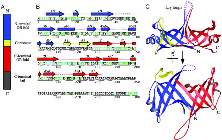
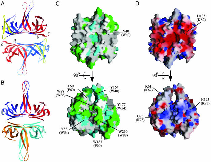
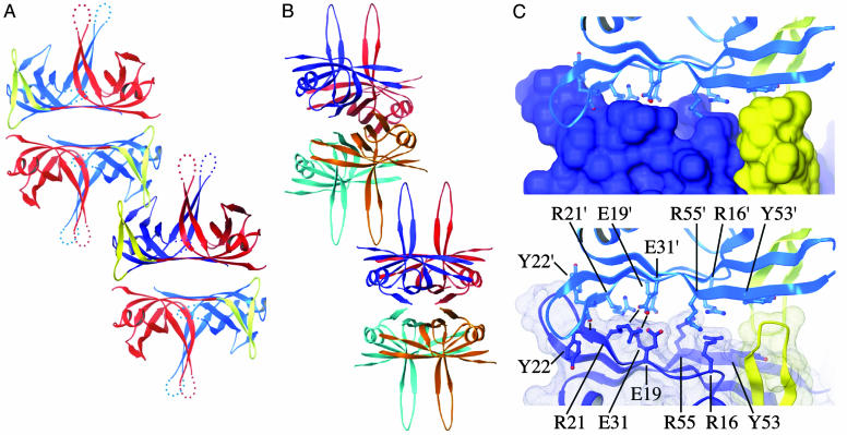
Single-stranded DNA (ssDNA)-binding (SSB) proteins are uniformly required to bind and protect single-stranded intermediates in DNA metabolic pathways. All bacterial and eukaryotic SSB proteins studied to date oligomerize to assemble four copies of a conserved domain, called an oligonucleotide/oligosaccharide-binding (OB) fold, that cooperate in nonspecific ssDNA binding. The vast majority of bacterial SSB family members function as homotetramers, with each monomer contributing a single OB fold. However, SSB proteins from the Deinococcus-Thermus genera are exceptions to this rule, because they contain two OB folds per monomer. To investigate the structural consequences of this unusual arrangement, we have determined a 1.8-A-resolution x-ray structure of Deinococcus radiodurans SSB. The structure shows that D. radiodurans SSB comprises two OB domains linked by a beta-hairpin motif. The protein assembles a four-OB-fold arrangement by means of symmetric dimerization. In contrast to homotetrameric SSB proteins, asymmetry exists between the two OB folds of D. radiodurans SSB because of sequence differences between the domains. These differences appear to reflect specialized roles that have evolved for each domain. Extensive crystallographic contacts link D. radiodurans SSB dimers in an arrangement that has important implications for higher-order structures of the protein bound to ssDNA. This assembly utilizes the N-terminal OB domain and the beta-hairpin structure that is unique to Deinococcus and Thermus species SSB proteins. We hypothesize that differences between D. radiodurans SSB and homotetrameric bacterial SSB proteins may confer a selective advantage to D. radiodurans cells that aids viability in environments that challenge genomic stability.


Figures

Fig. 1.
Fig. 1.
Structure of D. radiodurans SSB. (A) Schematic diagram of D. radiodurans SSB primary structure. Two OB folds are present in each D. radiodurans SSB monomer: one is N-terminal (blue, residues 1–108), and one is C-terminal (red, residues 129–233). These folds are linked by a connector peptide (yellow, residues 109–128). The C-terminal OB fold is followed by a flexible tail sequence (gray, residues 234–301). (B) Secondary structure of D. radiodurans SSB. The D. radiodurans SSB protein sequence is colored to indicate residues that are highly similar (teal) or identical (green) to E. coli SSB (28). Underlined residues are conserved with SSB proteins from T. aquaticus and T. thermophilus (13). Helices (boxes) and β-strands (arrows) are shown above the sequence (colored as in A) and labeled as for E. coli SSB (28) but with identifiers that indicate whether the element comprises part of the N-terminal (N) or the C-terminal (C) OB fold. β-strands in the connector region are labeled β1or β2 to distinguish them from elements in either OB fold. Dotted lines indicate regions for which electron density was not observed. Electron density was also absent for the C-terminal tail. (C) Orthogonal views of a ribbon diagram of the crystal structure of D. radiodurans SSB. The structure is colored as in A. Dotted lines indicate regions that could not be modeled because of a lack of electron density. The L45 loop region is indicated. Ribbon diagrams were rendered by using ribbons (33).
Fig. 2.
Fig. 2.
Dimeric structure of D. radiodurans SSB. (A) Ribbon diagram of the D. radiodurans SSB dimer. Coloring is as in Fig. 1 A for Upper and is shown in paler colors for Lower to aid in distinguishing between the two protomers. (B) Ribbon diagram of the E. coli SSB tetramer (28). Each protomer is colored differently (red, blue, orange, and teal). (C) Orthogonal views of the surface conservation of D. radiodurans SSB. The dimeric protein surface is colored as in Fig. 1B by its conservation with E. coli SSB. Structurally similar residues present at the positions of four key aromatic residues in E. coli SSB are indicated by their single-letter code with the equivalent E. coli residues indicated in parentheses. Upper is the same orientation as in A. (D) Orthogonal views of the surface potential of D. radiodurans SSB. The dimeric protein surface is colored by its electrostatic surface potential at ±6 kBT/e for positive (blue) or negative (red) charge potential by using the program grasp (34). Positions of several residues in D. radiodurans SSB are indicated in single-letter code, and electropositive DNA-binding residues in structurally conserved locations in E. coli SSB are indicated in parentheses. Upper is the same orientation as in A.
Fig. 3.
Fig. 3.
Higher-order assembly of D. radiodurans SSB. (A) Ribbon diagram of two crystallographically related D. radiodurans SSB dimers. One monomer is colored as in Fig. 1 A, with the other three shown in paler colors to aid in visualization. The N-terminal OB domains and β-hairpin connectors of the two central monomers interlock to form an extensive interaction surface. (B) Ribbon diagram of two crystallographically related E. coli SSB tetramers (28). Monomers are colored as in Fig. 2B. Interaction between tetramers is mediated by the L45 loops of two monomers. (C) Close-up view of the interaction surface between D. radiodurans SSB dimers. (Upper) The surface of one D. radiodurans SSB dimer is enveloped by its crystallographically related contact partner. Surfaces and ribbons are colored as in Fig. 1C, indicating that the interaction is mediated exclusively by the N-terminal OB domains and β-hairpin connectors of the adjacent protomers. (Lower) Residues that mediate the protein–protein contact (R21, Y22, E31, and Y53) and unpaired charged residues (R16, E19, and R55) are indicated. Residues labeled with a prime symbol are from the upper molecule.

References

    1. Lohman, T. M. & Ferrari, M. E. (1994) Annu. Rev. Biochem. 63, 527–570. - PubMed
    1. Mushegian, A. R. & Koonin, E. V. (1996) Proc. Natl. Acad. Sci. USA 93, 10268–10273. - PMC - PubMed
    1. Murzin, A. G. (1993) EMBO J. 12, 861–867. - PMC - PubMed
    1. Suck, D. (1997) Nat. Struct. Biol. 4, 161–165. - PubMed
    1. Shamoo, Y., Friedman, A. M., Parsons, M. R., Konigsberg, W. H. & Steitz, T. A. (1995) Nature 376, 362–366. - PubMed

Publication types

MeSH terms

LinkOut - more resources