. 2004 Jun;164(6):2013-25.
doi: 10.1016/S0002-9440(10)63761-3.

Plasmodium falciparum-infected erythrocytes adhere both in the intervillous space and on the villous surface of human placenta by binding to the low-sulfated chondroitin sulfate proteoglycan receptor

Affiliations

Plasmodium falciparum-infected erythrocytes adhere both in the intervillous space and on the villous surface of human placenta by binding to the low-sulfated chondroitin sulfate proteoglycan receptor

Arivalagan Muthusamy et al. Am J Pathol. 2004 Jun.

Abstract

In pregnant women infected with Plasmodium falciparum, the parasite-infected red blood cells (IRBCs) sequester in the placenta through chondroitin 4-sulfate (C4S)-mediated adherence. The pattern of IRBC adherence in P. falciparum-infected placenta has been controversial. Moreover, the identity of the chondroitin sulfate proteoglycan (CSPG) receptor, that mediates IRBC adherence, and its location in the placenta have not been established. This study, using immunohistochemical techniques, clearly shows, for the first time, that the low-sulfated CSPGs of the placenta are localized predominantly in the intervillous space. Ex vivo IRBC adherence analyses demonstrate that the IRBCs are adhered to the CSPG receptors in the placenta in a C4S-dependent manner. This IRBC binding pattern was similar to that observed in P. falciparum-infected placentas. These data and the results of dual-fluorescence staining of the endogenous RBCs and syncytiotrophoblasts, and co-localization of CSPG and IRBC adherence unequivocally establish that the low-sulfated CSPGs are the major natural receptors for IRBC adherence in the placenta. Further, it was found that IRBCs adhere mainly in the intervillous space and also at significant levels to the syncytiotrophoblasts. Finally, the ex vivo IRBC adherence method described herein provides a reliable procedure for future studies for the assessment of the efficacy of C4S inhibitors and adhesion inhibitory antibodies.


Figures

Figure 1
Figure 1
Enzyme-linked immunosorbent assay of antisera against placental proteoglycans. The purified low-sulfated CSPGs and DS/CSPG were coated onto 96-well microtiter plates at 20 μg/ml and 10 μg/ml, respectively. The wells were blocked with 0.5% casein in TBST, pH 7.5, and then incubated with either antisera or preimmune sera serially diluted in TBST containing 0.5% casein. After washing the plates, the bound antibodies were detected with horseradish peroxidase-conjugated goat anti-rabbit IgG using ABTS substrate. The assays were performed two times each in duplicates, and average OD values plotted. A: Antisera against the placental low-sulfated CSPGs before (•) and after (○) treatment of the CSPGs with chondroitinase ABC, and preimmune sera after treatment of the low-sulfated CSPGs with chondroitinase ABC (▴). B: Antisera against DS/CSPG before (•) and after (○) treatment of DS/CSPG with chondroitinase ABC, and preimmune sera after treatment of DS/CSPG with chondroitinase ABC (▴). In both cases the reactivities of preimmune sera before treatment with chondroitinase ABC were also at the background levels (not shown).
Figure 2
Figure 2
SDS-PAGE and Western blot analysis of the low-sulfated CSPGs and DS/CSPG purified from human placenta. A: Alcian Blue followed by silver staining of 4 to 15% gradient gel electrophoresed with the purified low-sulfated CSPGs and tissue matrix DS/CSPG before (lanes 1 and 3, respectively) and after (lanes 2 and 4, respectively) treatment with chondroitinase ABC. B: Immunoblots of the low-sulfated CSPGs and DS/CSPG before (lanes 1 and 3, respectively) and after (lanes 2 and 4, respectively) treatment with chondroitinase ABC using antibodies against the placental low-sulfated CSPGs. C: Immunoblots of the DS/CSPG and the low-sulfated CSPGs before (lanes 1 and 3, respectively) and after (lanes 2 and 4, respectively) treatment with chondroitinase ABC using antibodies specific to DS/CSPG. In all of the panels, each lane was loaded with 15 μg of proteoglycans. The positions (kd) of the molecular mass marker proteins are indicated on the right. Note: The arrow on the right indicates the position of chondroitinase ABC; the immunoreactivity of which is most likely because of the natural exposure of rabbits to the enzyme-producing bacteria.
Figure 3
Figure 3
Immunohistochemical localization of the CSPGs of human placenta. The placental tissue sections were immunostained with either polyclonal antibodies against the placental proteoglycan core proteins (B to D and F to H) or monoclonal antibodies specific to the Δdi-4S (I to L) and-Δdi-6S (M to P) moieties in the core proteins formed by the chondroitinase ABC treatment of the proteoglycans. A: Only background level of staining was observed with the preimmune serum from the rabbit used for raising antibodies against the placental low-sulfated CSPGs. B to D: Antibodies against the low-sulfated CSPGs stained fibrous, filamentous materials and fibrinoid deposits in the intervillous space. The fibrous projections from the syncytiotrophoblast layers were also stained. E: Preimmune serum from the rabbit used for raising antibodies against DS/CSPG showed no staining. F to H: Antibodies against the placental DS/CSPG stained strongly the perivascular regions of blood vessels and stroma. I: Anti-Δdi-4S monoclonal antibody did not stain tissue sections not treated with chondroitinase ABC. J to L: Anti-Δdi-4S monoclonal IgG moderately stained the matrix-like material of the intervillous space and strongly stained the perivascular regions of blood vessels, extending to the stroma in the chondroitinase ABC-treated tissue sections. M: Anti-Δdi-6S monoclonal antibody did not stain the tissue sections not treated with chondroitinase ABC. N to P: Anti-Δdi-6S monoclonal IgM stained strongly the stroma in the chondroitinase ABC-treated tissue sections. C represents the enlarged area marked in B. Ivs, intervillous space; Syn, syncytiotrophoblasts; RBC, red blood cells; Fbd, fibrinoid deposits; Fbv, fetal blood vessel; St, stromal tissue. Arrows indicate the staining of the fibrous filament-like materials in the intervillous space. Original magnifications, ×100 (A, B, D–P).
Figure 4
Figure 4
Ex vivo adherence of P. falciparum IRBCs to human placenta. The 3D7-C4S parasites or placental blood samples were prestained with SYBR Green and overlaid onto the placental tissue sections fixed with formaldehyde and glutaraldehyde followed by microwave treatment. The unbound IRBCs and RBCs were washed, and the sections were examined and photographed under fluorescence microscopy (A, C, E, G, I, and K) and light microscopy (B, D, F, H, J, and L). A and C: Fluorescent micrographs of the tissue sections showing SYBR Green-stained 3D7-C4S IRBCs densely adhered in the intervillous space and moderately adhered to the syncytiotrophoblasts. B and D: Light micrographs of fields corresponding to A and C, respectively. E and G: Fluorescent micrographs of the tissue sections showing SYBR Green-stained placental IRBCs adhered in the intervillous space and to syncytiotrophoblast cell layer. F and H: Light micrographs of fields corresponding to E and G, respectively. I: Fluorescent micrograph of the chondroitinase ABC-treated tissue sections overlaid with the SYBR Green-stained IRBCs. J: Light micrograph of the field corresponding to I; the RBCs (arrowhead) present in J were because of those present in the fixed tissue section (also see Figure 5). K: Fluorescent micrograph of the tissue sections overlaid with the SYBR Green-stained IRBCs preincubated with 80 μg/ml of C4S. L: Light micrograph of the field corresponding to K; as in J, the observed RBCs (arrowhead) were because of those present in the fixed tissue section (also see Figure 5). Ivs, intervillous space; Syn, syncytiotrophoblasts; Fbv, fetal blood vessels; St, stromal tissue. Arrows indicate the IRBCs; arrowheads indicate the RBCs. Original magnifications, ×40.
Figure 5
Figure 5
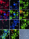
Co-localization of the low-sulfated CSPGs and IRBC adherence in the placenta, and dual-fluorescence staining of syncytiotrophoblasts and endogenous RBCs in the placenta. Placental tissue sections were stained with DAPI and anti-CSPG or anti-glycophorin A and B antibodies, and then adherence with SYBR Green-stained IRBCs performed as described in Figure 4. A to C: Fluorescent micrographs of the tissue sections stained with the antibodies against the placental low-sulfated CSPGs (A) and with DAPI (B) followed by adherence with SYBR Green-stained IRBCs (C). D to F: Merged pictures of A and B (D), B and C (E), and A and C (F). G to I: Fluorescent micrograph of the tissue sections stained with the monoclonal antibodies specific to human glycophorin A and B (G) and with DAPI (H) followed by adherence with SYBR Green-stained IRBCs (I). J and K: Merged pictures of H and I (J) and G and I (K). L: Light micrograph of tissue sections shown in G to K. Ivs, intervillous space; Syn, syncytiotrophoblasts; Fbv, fetal blood vessels; St, stromal tissue. Arrows indicate the IRBCs; arrowheads indicate the RBCs. Original magnifications, ×40.
Figure 6
Figure 6
Adherence of IRBCs in P. falciparum-infected human placenta. The P. falciparum-infected placental tissue was fixed with 10% neutral buffered formalin, sectioned, stained with H&E, and photographed under light microscopy. Massive adherence of IRBCs (those RBCs with stained parasite inside) in the intervillous space, as well as significant level of adherence to syncytiotrophoblast lining in four different infected placentas (A to D) with 16% (A), 41% (B), 46% (C), and 50% (D) placental parasitemia, respectively. Arrows indicate the IRBCs; arrowheads indicate the RBCs. Original magnifications, ×40.

References

    1. Trigg T, Kondrachine AV. The current global malaria situation. Sherman IW, editor. Washington DC: ASM Press,; Malaria—Parasite Biology, Pathogenesis, and Protection. 1998:pp 11–22.
    1. Snow RW, Craig M, Diechmann U, Marsh K. Estimating mortality, morbidity and disability due to malaria among Africa’s non-pregnant population. Bull WHO. 1999;77:624–640. - PMC - PubMed
    1. Greenwood B, Mutabingwa T. Malaria in 2002. Nature. 2002;415:670–672. - PubMed
    1. Miller LH, Baruch DI, Marsh K, Doumbo OK. The pathogenic basis of malaria. Nature. 2002;415:673–679. - PubMed
    1. Pasloske BL, Howard RJ. Malaria, the red cell, and the endothelium. Annu Rev Med. 1994;45:283–295. - PubMed

Publication types

Substances

LinkOut - more resources