. 2004 Jun;164(6):2089-99.
doi: 10.1016/S0002-9440(10)63767-4.

Macrophage/microglial accumulation and proliferating cell nuclear antigen expression in the central nervous system in human immunodeficiency virus encephalopathy

Affiliations

Macrophage/microglial accumulation and proliferating cell nuclear antigen expression in the central nervous system in human immunodeficiency virus encephalopathy

Tracy Fischer-Smith et al. Am J Pathol. 2004 Jun.

Abstract

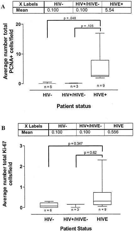
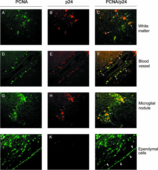
This study was performed to quantitate and characterize the mononuclear phagocytes (MPs) in human immunodeficiency virus encephalopathy (HIVE) by immunohistochemistry in an effort to gain insights into potential mechanisms of central nervous system (CNS) accumulation. Single- and double-labeled studies using antibodies against CD14, CD16, CD68, proliferating cell nuclear antigen (PCNA), Ki-67, von Willebrand factor, and HIV-1 p24 were performed using brain tissue from patients with HIVE, HIV-1 infection without encephalitis, and seronegative controls. A substantial increase in MPs was observed in CNS tissue from patients with HIVE, relative to seronegative controls and patients with acquired immune deficiency syndrome but without encephalitis, as determined by CD68 and CD16 immunohistochemistry. A large proportion of CD16+ MPs in HIVE CNS tissue were PCNA+, but do not appear to be proliferating, based on limited Ki-67 positivity. Although virtually all cells positive for HIV-1 p24 were PCNA+, there were many PCNA+ cells where HIV-1 p24 expression was not detected. PCNA positivity was also observed in some endothelial cells and ependymal cells in HIVE CNS. Our results would support a role for HIV-1-induced alterations in MP trafficking and homeostasis in the pathogenesis of HIVE.


Figures

Figure 1
Figure 1
CD68, CD14, and CD16 analysis of variance box plots. A significant number of total brain MPs (A), as determined by CD68 positivity, were observed in HIVE when compared to HIV-1-infected individuals without dementia (P = 0.016) and seronegative controls (P = 0.002). HIV+/HIVE− versus HIV− was not statistically significant (P = 0.861). Similarly, parenchymal CD68+ MPs (B) demonstrated statistical significance between HIVE and HIV-1 without dementia (P = 0.003) and HIVE and seronegative individuals (P = 0.001). CD16+ cells in HIVE parenchyma were also present in statistically greater numbers, as compared to HIV+/HIVE− (P = 0.03), as well as seronegative controls (P = 0.005) (C), however, CD14+ parenchymal cells did not increase significantly between controls and HIVE brains (D). Results for all comparisons were verified by the Tukey-Kramer comparison of means posttest.
Figure 2
Figure 2
PCNA and CD16 immunohistochemistry. A and E: Normal brain; B and F: HIV-1 without encephalopathy; C, G, and I: HIVE; D, H, and J: CD16 immunohistochemistry in HIVE brain tissue. The top row (A–D) illustrates white matter. E to H: Blood vessels. I and J: Microglial nodules in the vicinity of blood vessels (arrows). In HIVE, PCNA+ cells were observed in white matter, perivascularly, and within microglial nodules (C, G, and I, arrows). A small number of PCNA+ cells were observed perivascularly in patients with HIV-1 infection but without dementia (F, arrow). Some cells observed within blood vessels, not associated with brain tissue, were also PCNA+ in HIV+/HIVE− and HIVE+ brain tissue (F and I, arrowheads). CD16+ cells observed in white matter of patients with HIVE had a ramified morphology (D, arrows) and were also seen perivascularly and within microglial nodules in HIVE (H and J). Original magnifications, ×40.
Figure 3
Figure 3
Ki-67 immunohistochemistry. A and D: Normal brain; B and E: HIV-1 without encephalopathy; C, F, and H: HIVE; G: small intestine from a seronegative individual. The top row (A–C) illustrates white matter. D–F: Blood vessels. H: A multinucleated giant cell within a microglial nodule. PCNA+ brain tissue from patients with HIVE showed only occasional Ki-67-labeled nuclei, as demonstrated in C (white matter) and F (blood vessel). H: Multinucleated giant cells did not show Ki-67 positivity. Rare positivity was observed in white matter of patients with HIV-1 infection without dementia (B) and seronegative controls (A). E: Limited positive cells were also located perivascularly in HIV+/HIVE− brain tissue. G: Autopsy intestine from a seronegative individual shows abundant nuclear Ki-67 positivity with the CSA technique. Original magnifications: ×40 (A–G); ×100 (H).
Figure 4
Figure 4
PCNA and Ki-67 analysis of variance box plots. A: The total number of PCNA+ cells was significantly increased in HIVE relative to seronegative controls using the Tukey-Kramer test for multiple comparisons (P = 0.048). Although the difference between HIV-1/AIDS and HIVE did not reach significance (P = 0.105), the number of patients is small with a large SD because of local variations in staining within specimens. After dividing the patients into HIVE− and HIVE+, the encephalopathy group demonstrated increased PCNA positivity relative to the patients without encephalopathy (P = 0.01) by Student’s t-test, assuming unequal variances. In contrast, the small increase in the number of Ki-67-positive cells in HIVE relative to HIV-1/AIDS and seronegative controls, was not significant (B) and did not reach statistical significance after combining the HIV-1/AIDS and control groups into HIVE− and HIVE+ (P = 0.089). Results for all comparisons were verified by the Tukey-Kramer comparison of means posttest.
Figure 5
Figure 5
PCNA/CD16 immunohistochemistry. All panels show PCNA/CD16 double immunohistochemistry on brain tissue from a patient with HIVE (HIVE 08). In HIVE, PCNA (brown) and CD16 (pink) double-positive cells were observed in white matter (A and B), around blood vessels (C), and within microglial nodules (D and E). B: Inset of A. E: Inset of D. Arrows point to cells with co-localized PCNA and CD16 expression. Some PCNA+ cells in white matter and around blood vessels did not express detectable levels of CD16 (A–C, arrowheads). Multinucleated giant cells, not associated with microglial nodules, were also positive for both antigens (F). Original magnifications: ×40 (A–E); ×100 (F).
Figure 6
Figure 6
PCNA/von Willebrand factor immunohistochemistry. All panels show PCNA/von Willebrand factor double immunohistochemistry on brain tissue from a patient with HIVE (HIVE 08). In brain tissue from patients with HIVE, several endothelial cells, which are positive for von Willebrand Factor (pink), were also PCNA-positive (brown). Arrows indicate double-positive cells. Original magnifications: ×40 (A); ×100 (B).
Figure 7
Figure 7
PCNA/p24 immunofluorescence. All panels show PCNA/p24 immunofluorescence on brain tissue from a patient with HIVE (HIVE 08). fluorescein isothiocyanate-labeled PCNA+ cells (green) were observed in white matter (A), around blood vessels (D), within microglial nodules (G), and in ependymal cells (J). Texas Red-labeled HIV-1 p24+ cells (red) were observed in white matter (B), around blood vessels (E), and within microglial nodules (H), but not in ependymal cells (K). Approximately half of the PCNA+ cells were also p24+ (yellow) (C, F, and I). Arrows point to cells that are positive for PCNA and p24. Some multinucleated giant cells, not associated with nodules, in white matter were also positive for both antigens (C, short-tailed arrows). Although virtually all p24+ cells were PCNA+, many PCNA+ cells were negative for HIV-1 p24, including cells located in white matter, perivascularly, and within microglial nodules (C, F, and I, arrowheads), as well as all ependymal cells (L, arrowheads). Original magnifications, ×40.

References

    1. Sacktor N, Lyles RH, Skolasky R, Kleeberger C, Selnes OA, Miller EN, Becker JT, Cohen B, McArthur JC. HIV-associated neurologic disease incidence changes: Multicenter AIDS Cohort Study, 1990–1998. Neurology. 2001;56:257–260. - PubMed
    1. McArthur JC, Hoover DR, Bacellar H, Miller EN, Cohen BA, Becker JT, Graham NM, McArthur JH, Selnes OA, Jacobson LP, Visscher BR, Concha M, Saah S. Dementia in AIDS patients: incidence and risk factors. Multicenter AIDS Cohort Study. Neurology. 1993;43:2245–2252. - PubMed
    1. Vazeux R, Lacroix-Ciaudo C, Blanche S, Cumont MC, Henin D, Gray F, Boccon-Gibod L, Tardieu M. Low levels of human immunodeficiency virus replication in the brain tissue of children with severe acquired immunodeficiency syndrome encephalopathy. Am J Pathol. 1992;140:137–144. - PMC - PubMed
    1. Rostad SW, Sumi SM, Shaw CM, Olson K, McDougall JK. Human immunodeficiency virus (HIV) infection in brains with AIDS-related leukoencephalopathy. AIDS Res Hum Retroviruses. 1987;3:363–373. - PubMed
    1. Pumarola-Sune T, Navia BA, Cordon-Cardo C, Cho ES, Price RW. HIV antigen in the brains of patients with the AIDS dementia complex. Ann Neurol. 1987;21:490–496. - PubMed

Publication types