Review
. 2004 Jan;3(1):105-20.
doi: 10.1016/j.arr.2003.08.005.

Infectious agents and age-related neurodegenerative disorders

Affiliations
Review

Infectious agents and age-related neurodegenerative disorders

Mark P Mattson. Ageing Res Rev. 2004 Jan.

Erratum in

  • Ageing Res Rev. 2004 Apr;3(2):349

Abstract

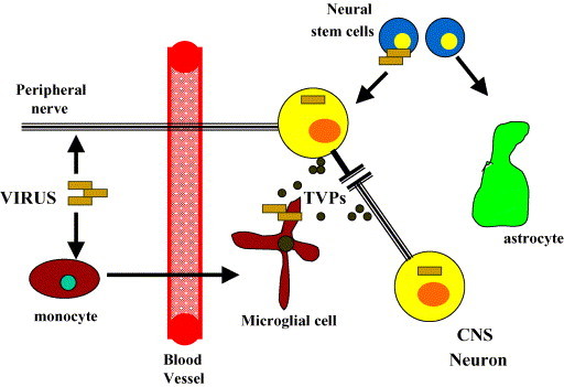
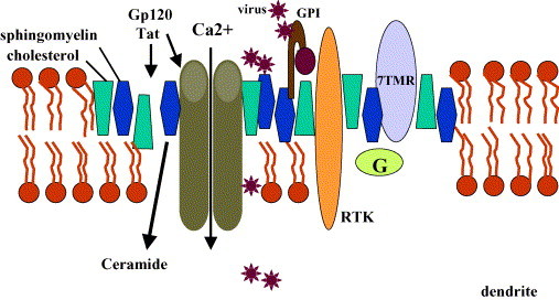
chlamdAs with other organ systems, the vulnerability of the nervous system to infectious agents increases with aging. Several different infectious agents can cause neurodegenerative conditions, with prominent examples being human immunodeficiency virus (HIV-1) dementia and prion disorders. Such infections of the central nervous system (CNS) typically have a relatively long incubation period and a chronic progressive course, and are therefore increasing in frequency as more people live longer. Infectious agents may enter the central nervous system in infected migratory macrophages, by transcytosis across blood-brain barrier cells or by intraneuronal transfer from peripheral nerves. Synapses and lipid rafts are important sites at which infectious agents may enter neurons and/or exert their cytotoxic effects. Recent findings suggest the possibility that infectious agents may increase the risk of common age-related neurodegenerative disorders such as Alzheimer's disease (AD) and Parkinson's disease (PD), amyotrophic lateral sclerosis (ALS) and stroke. While scenarios can be envisioned whereby viruses such as Chlamydia pneumoniae, herpes simplex and influenza promote damage to neurons during aging, there is no conclusive evidence for a major role of these pathogens in neurodegenerative disorders. In the case of stroke, blood vessels may be adversely affected by bacteria or viruses resulting in atherosclerosis.


Figures

Fig. 1
Fig. 1
Mechanisms whereby viruses infect the CNS and cause neuronal dysfunction and death. Two routes of entry of viruses into the CNS are by infection of cells that cross the blood–brain barrier (e.g. cells of the monocyte–macrophage/microglia lineage) and intra- and transneuronal transfer from peripheral neurons. Microglia and neural stem cells appear to be cell types in which viruses may replicate at a high rate; viruses may also infect neurons and astrocytes, but may not replicate in such postmitotic cells. Viruses can produce toxic viral proteins (TVPs) with the HIV-1 proteins gp120 and Tat being exemplary.
Fig. 2
Fig. 2
Roles of membrane lipid rafts in infections of the central nervous system. Lipid rafts are microdomains of the plasma membrane in which the outer leaflet of the phospholipid bilayer contains relatively high levels of cholesterol and sphingomyelin. A variety of receptors and associated signal transduction proteins (receptor tyrosine kinases, RTK; seven transmembrane receptors, 7TMR coupled to GTP-binding proteins), and ion channels are concentrated in lipid rafts. Viruses may gain entry to cells by binding to proteins (e.g. GPI-anchored receptors) or lipids in lipid rafts. Lipid rafts may also be regions of membranes that are damaged by cytotoxic viral proteins such as the HIV-1 proteins gp120 and Tat.
Fig. 3
Fig. 3
Similarities in the neurotoxic actions of pathogenic forms of prion proteins and amyloid β-peptide (Aβ). Normal forms of Aβ (soluble Aβ) and prion proteins do not self aggregate, whereas in prion disorders and Alzheimer’s disease the proteins generate reactive oxygen species (ROS) and acquire abnormal conformations. Genetic factors (e.g. mutations in the genes encoding the prion protein or Aβ) and environmental factors (e.g. high caloric intake or folate deficiency) and the aging process may cause or promote formation of pathogenic forms of Aβ and PrPsc. During the process of peptide oligomer and fibril formation, Aβ and PrPsc induce membrane-associated oxidative stress and disrupt membrane ion transporter and channel functions resulting in synaptic and mitochondrial dysfunction and apoptosis. It has recently become evident that autoantibodies against abnormal conformations of Aβ and PrPsc can be generated by the immune systems in patients and in subjects immunized with Aβ or prion peptides. The autoantibodies may promote clearance of the endogenous pathogenic proteins from the brain and/or they may enhance the neurotoxicity of Aβ or PrPsc.
Fig. 4
Fig. 4
Cellular and molecular mechanisms responsible for motor dysfunction in Parkinson’s disease (PD) and amyotrophic lateral sclerosis (ALS). Some rare cases of PD and ALS are caused by mutations. Three genes have been identified in which mutations cause early onset inherited PD (Parkin, α-synuclein and DJ-1) and mutations in two genes (Cu/Zn-SOD and Alsin) are known to cause ALS. Insight into the molecular and cellular abnormalities that lead to the dysfunction and death of midbrain dopaminergic neurons in PD and of motor neurons in ALS has come from studies of cultured cells and transgenic mice expressing the disease-causing human genes. In the case of PD, each of the mutations has been linked to abnormal ubiquitin/proteasome-mediated proteolysis, cellular oxidative and metabolic stress and triggering of apoptosis. In the case of ALS, Cu/Zn-SOD mutations cause oxidative stress and disrupt cellular calcium homeostasis. The aging process and environmental factors may perturb the same or similar regulatory systems that are adversely affected by genetic mutations. Synapses are particularly vulnerable to genetic, aging and environmental factors, and are sites where excitotoxic and apoptotic cascades that cause the death of the neurons are initiated.

Similar articles

Cited by

References

    1. Antezana D.F., Clatterbuck R.E., Alkayed N.J., Murphy S.J., Anderson L.G., Frazier J., Hurn P.D., Traystman R.J., Tamargo R.J. High-dose ibuprofen for reduction of striatal infarcts during middle cerebral artery occlusion in rats. J. Neurosurg. 2003;98:860–866. - PubMed
    1. Balin B.J., Appelt D.M. Role of infection in Alzheimer’s disease. J. Am. Osteopath. Assoc. 2001;101:S1–6. - PubMed
    1. Balin B.J., Gerard H.C., Arking E.J., Appelt D.M., Branigan P.J., Abrams J.T., Whittum-Hudson J.A., Hudson A.P. Identification and localization of Chlamydia pneumoniae in the Alzheimer’s brain. Med. Microbiol. Immunol. (Berl.) 1998;187:23–42. - PubMed
    1. Berger J.R., Arendt G. HIV dementia: the role of the basal ganglia and dopaminergic systems. J. Psychopharmacol. 2000;14:214–221. - PubMed
    1. Bruce A.J., Boling W., Kindy M.S., Peschon J., Kraemer P.J., Carpenter M.K., Holtsberg F.W., Mattson M.P. Altered neuronal and microglial responses to brain injury in mice lacking TNF receptors. Nat. Med. 1996;2:788–794. - PubMed

MeSH terms