

doi: 10.1242/dev.01158.
Epub 2004 May 26.
Development of the mammary gland requires DGAT1 expression in stromal and epithelial tissues
Affiliations
- PMID: 15163627
- PMCID: PMC2775443
- DOI: 10.1242/dev.01158
Item in Clipboard
Development of the mammary gland requires DGAT1 expression in stromal and epithelial tissues
Development.
2004 Jul.
Abstract
Mammary gland development is a complex process that is dependent on interactions between the developing mammary epithelium and the surrounding stromal tissues. We show that mice lacking the triglyceride synthesis enzyme acyl CoA:diacylglycerol transferase 1 (DGAT1) have impaired mammary gland development, characterized by decreased epithelial proliferation and alveolar development, and reduced expression of markers of functional differentiation. Transplantation studies demonstrate that the impaired development results from a deficiency of DGAT1 in both the stromal and epithelial tissues. Our findings are the first to link defects in stromal lipid metabolism to impaired mammary gland development.
Figures
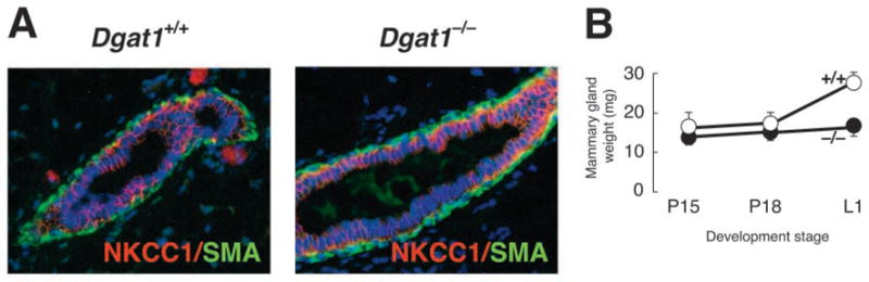
Characterization of mammary glands of virgin and pregnant Dgat1−/− mice. (A) Immunohistochemistry of mammary glands from 8-week-old virgin female Dgat1+/+ and Dgat1−/− mice. After fixation, mammary tissue was embedded in paraffin, sectioned, and stained with antibodies specific for the ductal cell marker NKCC1 (red) and smooth muscle actin (SMA, green). (B) Weights of inguinal mammary glands from wild-type and Dgat1−/− mice during pregnancy.
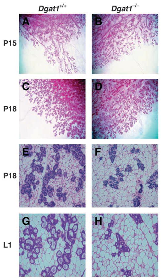
Reduced alveolar development in mammary glands of Dgat1−/− mice. Whole mounts (A–D) and histological sections (E–H) of mammary glands of wild-type and Dgat1−/− mice at P15, P18 and L1. At least five mice of each genotype were analyzed. Representative sections are shown.
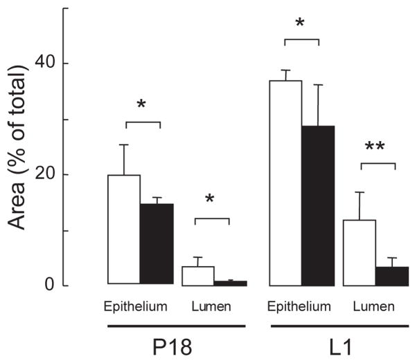
Reduced alveolar development in mammary glands of Dgat1−/− mice. Epithelial and luminal areas were quantified in Dgat1+/+ (white bars) and Dgat1−/− (black bars) mammary glands using digitalized images of the histology sections at P18 and L1. Area was quantified in five random fields. n=4 Dgat1+/+ mice; n=5 Dgat1−/− mice. *P<0.05; **P<0.01
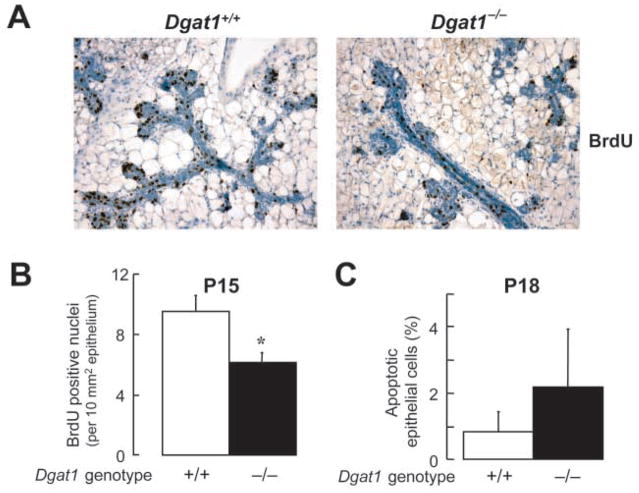
(A) Bromodeoxyuridine (BrdU) immunostaining. Pregnant (P15) mice were injected with BrdU 2 hours before mammary glands were removed. Tissues were fixed, embedded and sectioned, and proliferating nuclei were detected by BrdU immunostaining (dark brown staining). (B) Quantification of BrdU-positive nuclei. Epithelial areas were calculated using images of histological sections (see Materials and methods), and positive nuclei from five random fields were counted and expressed per unit of area (*P<0.05, n=4 mice per genotype). The expression of BrdU-positive nuclei as a percentage of total epithelial nuclei showed a similar reduction in proliferating nuclei (9.76±1.02% for Dgat1−/− versus 17.82±0.3% for wild type, P=0.002, n=3). (C) Similar levels of apoptosis were seen in mammary glands of wild-type and Dgat1−/− mice at P18. Apoptotic epithelial cells were detected by TUNEL staining and were counted in 10 random fields. n=5 mice per genotype.
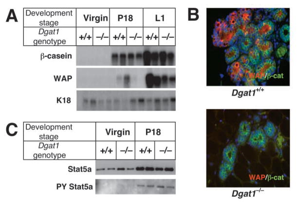
Impaired differentiation of mammary epithelium in Dgat1−/− mice. (A) Reduced expression of markers of mammary epithelial differentiation in Dgat1−/− mice. Total mammary gland RNA was extracted from wild-type and Dgat1−/− virgin mice, and at P18 and L1. Expression of the milk protein genes β-casein and Wap was analyzed by northern blotting. The expression of keratin 18 (K18) was used as a control for the content of RNA from mammary epithelium. (B) Reduced immunostaining of WAP in Dgat1−/− mammary glands. Immunostaining of β-catenin (β-cat) was used as control. (C) Similar STAT5A expression and phosphorylation were observed in wild-type and Dgat1−/− mammary glands. Glands from virgin mice and from mice at P18 were dissected, and homogenates were prepared. STAT5A was immunoprecipitated from these samples and analyzed by immunoblotting with antibodies that detect STAT5A or phosphorylated tyrosine residues (PY).
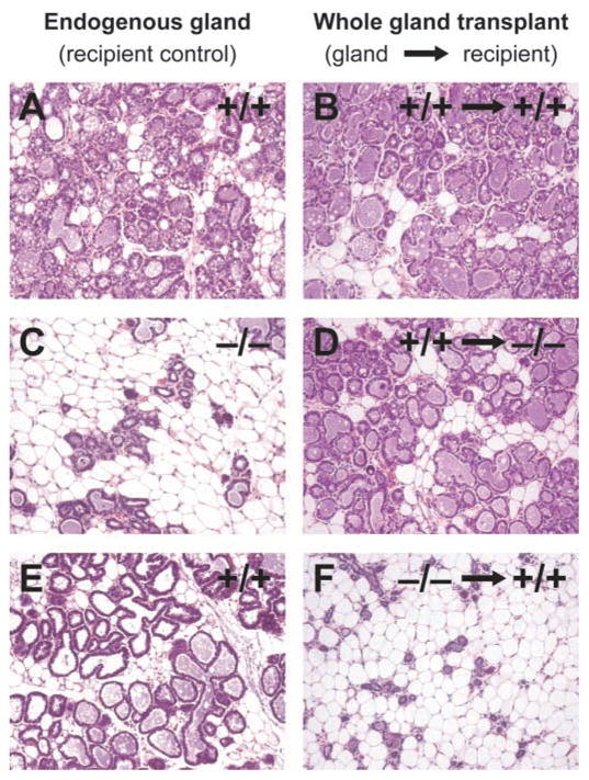
Whole mammary gland transplantations. Whole mammary glands were transplanted into the intrascapular region of recipient mice, and sections were analyzed at L1. In control experiments, whole wild-type mammary gland transplants (B) developed to the same extent as wild-type endogenous glands (A). Although control recipient Dgat1−/− glands failed to develop (C), wild-type whole mammary glands transplanted into the same Dgat1−/− recipients developed normally (D). In the converse experiment, control endogenous Dgat1+/+ glands developed normally (E), whereas Dgat1−/− mammary glands transplanted into the Dgat1+/+ recipients failed to develop (F).
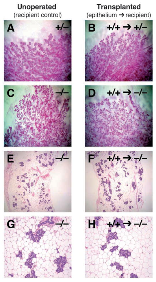
Transplantation of Dgat1+/+ mammary epithelium into Dgat1−/− recipient mice. Recipient mammary glands were cleared of endogenous epithelium and then implanted with donor epithelium. Recipient mice were mated, and epithelial morphology was analyzed at P18 by wholemounts (A-D) or histology (E-H). Shown are recipient (unoperated) control glands (A,C,E,G) and recipient glands that received transplanted epithelium (B,D,F,H). Control transplantations are shown for comparison (A,B).
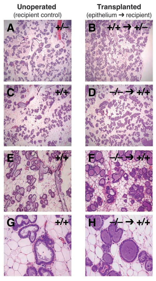
Transplantation of Dgat1−/− epithelium into Dgat1+/+ recipient mice. (A–H) After mammary epithelial transplantation, recipient mice were mated and epithelial morphology was analyzed by histology at L1. Shown are recipient (unoperated) control glands (A,C,E,G) and recipient glands that received transplanted epithelium (B,D,F,H). Control transplantations are shown for comparison (A,B).
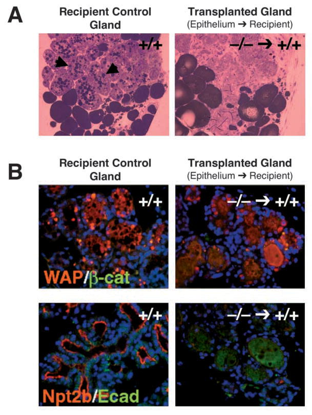
Impaired lactation and differentiation in Dgat1−/− mammary epithelium transplanted into wild-type recipient mammary glands. (A) Absence of lipid droplets in Dgat1−/− mammary epithelium transplanted into wild-type recipient mammary glands. Sections were stained with osmium tetroxide, which stains neutral lipids. Note the lipid droplets in the recipient control glands (arrowheads). (B) Reduced immunostaining of markers of epithelial differentiation in Dgat1−/− mammary epithelium transplanted into wild-type recipient mammary glands. Red fluorescence indicates the differentiation markers WAP and NPT2B. Green fluorescence indicates the control markers of epithelial cells, β-catenin (β-cat) and E-cadherin (Ecad).
References
-
- Bell RM, Coleman RA. Enzymes of glycerolipid synthesis in eukaryotes. Annu Rev Biochem. 1980;49:459–487. - PubMed
-
- Brindley DN. Metabolism of triacylglycerols. In: Vance DE, Vance JE, editors. Biochemistry of Lipids, Lipoproteins and Membranes. Amsterdam: Elsevier; 1991. pp. 171–203.
-
- Brisken C, Kaur S, Chavarria TE, Binart N, Sutherland RL, Weinberg RA, Kelly PA, Ormandy CJ. Prolactin controls mammary gland development via direct and indirect mechanisms. Dev Biol. 1999;210:96–106. - PubMed
Publication types
MeSH terms
Substances
Grants and funding
LinkOut - more resources
Full Text Sources
Molecular Biology Databases
