Response properties of whisker-related neurons in rat second somatosensory cortex
- PMID: 15163670
- PMCID: PMC2804247
- DOI: 10.1152/jn.00262.2004
Response properties of whisker-related neurons in rat second somatosensory cortex
Abstract
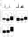
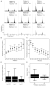
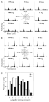
In addition to a primary somatosensory cortex (SI), the cerebral cortex of all mammals contains a second somatosensory area (SII); however, the functions of SII are largely unknown. Our aim was to explore the functions of SII by comparing response properties of whisker-related neurons in this area with their counterparts in the SI. We obtained extracellular unit recordings from narcotized rats, in response to whisker deflections evoked by a piezoelectric device, and compared response properties of SI barrel (layer IV) neurons with those of SII (layers II to VI) neurons. Neurons in both cortical areas have similar response latencies and spontaneous activity levels. However, SI and SII neurons differ in several significant properties. The receptive fields of SII neurons are at least five times as large as those of barrel neurons, and they respond equally strongly to several principal whiskers. The response magnitude of SII neurons is significantly smaller than that of neurons in SI, and SII neurons are more selective for the angle of whisker deflection. Furthermore, whereas in SI fast-spiking (inhibitory) and regular-spiking (excitatory) units have different spontaneous and evoked activity levels and differ in their responses to stimulus onset and offset, SII neurons do not show significant differences in these properties. The response properties of SII neurons suggest that they are driven by thalamic inputs that are part of the paralemniscal system. Thus whisker-related inputs are processed in parallel by a lemniscal system involving SI and a paralemniscal system that processes complimentary aspects of somatosensation.
Figures


Similar articles
-
Spatial gradients and inhibitory summation in the rat whisker barrel system.J Neurophysiol. 1996 Jul;76(1):130-40. doi: 10.1152/jn.1996.76.1.130. J Neurophysiol. 1996. PMID: 8836214
-
Intrinsic firing patterns and whisker-evoked synaptic responses of neurons in the rat barrel cortex.J Neurophysiol. 1999 Mar;81(3):1171-83. doi: 10.1152/jn.1999.81.3.1171. J Neurophysiol. 1999. PMID: 10085344
-
Sub- and suprathreshold receptive field properties of pyramidal neurones in layers 5A and 5B of rat somatosensory barrel cortex.J Physiol. 2004 Apr 15;556(Pt 2):601-22. doi: 10.1113/jphysiol.2003.053132. Epub 2004 Jan 14. J Physiol. 2004. PMID: 14724202 Free PMC article.
-
Differential origin of projections from SI barrel cortex to the whisker representations in SII and MI.J Comp Neurol. 2006 Oct 10;498(5):624-36. doi: 10.1002/cne.21052. J Comp Neurol. 2006. PMID: 16917827
-
Deconstructing the cortical column in the barrel cortex.Neuroscience. 2018 Jan 1;368:17-28. doi: 10.1016/j.neuroscience.2017.07.034. Epub 2017 Jul 21. Neuroscience. 2018. PMID: 28739527 Review.
Cited by
-
Sensory and decision-related activity propagate in a cortical feedback loop during touch perception.Nat Neurosci. 2016 Sep;19(9):1243-9. doi: 10.1038/nn.4356. Epub 2016 Jul 20. Nat Neurosci. 2016. PMID: 27437910 Free PMC article.
-
Layer-specific touch-dependent facilitation and depression in the somatosensory cortex during active whisking.J Neurosci. 2006 Sep 13;26(37):9538-47. doi: 10.1523/JNEUROSCI.0918-06.2006. J Neurosci. 2006. PMID: 16971538 Free PMC article.
-
Anatomically and functionally distinct thalamocortical inputs to primary and secondary mouse whisker somatosensory cortices.Nat Commun. 2020 Jul 3;11(1):3342. doi: 10.1038/s41467-020-17087-7. Nat Commun. 2020. PMID: 32620835 Free PMC article.
-
A Non-canonical Feedback Circuit for Rapid Interactions between Somatosensory Cortices.Cell Rep. 2018 May 29;23(9):2718-2731.e6. doi: 10.1016/j.celrep.2018.04.115. Cell Rep. 2018. PMID: 29847801 Free PMC article.
-
Bilateral plasticity of Vibrissae SII representation induced by classical conditioning in mice.J Neurosci. 2011 Apr 6;31(14):5447-53. doi: 10.1523/JNEUROSCI.5989-10.2011. J Neurosci. 2011. PMID: 21471380 Free PMC article.
References
-
- Alloway KD, Burton H. Homotypical ipsilateral cortical projections between somatosensory areas I and II in the cat. Neuroscience. 1985a;14:15–35. - PubMed
-
- Alloway KD, Burton H. Submodality and columnar organization of the second somatic sensory area in cats. Exp Brain Res. 1985b;61:128–140. - PubMed
-
- Armstrong-James M. The nature and plasticity of sensory processing within adult rat barrel cortex. In: Jones EG, Diamond IT, editors. Cerebral Cortex: The Barrel Cortex of Rodents. Vol. 11. New York: Plenum Press; 1995. pp. 333–373.
-
- Armstrong-James M, Fox K, Dasgupta A. Flow of excitation within rat barrel cortex on striking a single vibrissa. J Neurophysiol. 1992;68:1345–1358. - PubMed
-
- Brumberg JC, Pinto DJ, Simons DJ. Cortical columnar processing in the rat whisker-to-barrel system. J Neurophysiol. 1999;82:1808–1817. - PubMed
Publication types
MeSH terms
Grants and funding
LinkOut - more resources
Full Text Sources

