. 2004 May 26;24(21):4935-40.
doi: 10.1523/JNEUROSCI.0795-04.2004.

Firing mode-dependent synaptic plasticity in rat neocortical pyramidal neurons

Affiliations

Firing mode-dependent synaptic plasticity in rat neocortical pyramidal neurons

Barbara Birtoli et al. J Neurosci. .

Abstract

Pyramidal cells in the mammalian neocortex can emit action potentials either as series of individual spikes or as distinct clusters of high-frequency bursts. However, why two different firing modes exist is largely unknown. In this study, we report that in layer V pyramidal cells of the rat somatosensory cortex, in vitro associations of EPSPs with spike bursts delayed by +10 msec led to long-term synaptic depression (LTD), whereas pairings with individual action potentials at the same delay induced long-term potentiation. EPSPs were evoked extracellularly in layer II-III and recorded intracellularly in layer V neurons with the whole-cell or nystatin-based perforated patch-clamp technique. Bursts were evoked with brief somatic current injections, resulting in three to four action potentials with interspike frequencies of approximately 200 Hz, characteristic of intrinsic burst firing. Burst-firing-associated LTD (Burst-LTD) was robust over a wide range of intervals between -100 and +200 msec, and depression was maximal (approximately 50%) for closely spaced presynaptic and postsynaptic events. Burst-LTD was associative and required concomitant activation of low voltage-activated calcium currents and metabotropic glutamate receptors. Conversely, burst-LTD was resistant to blockade of NMDA receptors or inhibitory synaptic potentials. Burst-LTD was also inducible at already potentiated synapses. We conclude that intrinsic burst firing represents a signal for resetting excitatory synaptic weights.


Figures

Figure 1.
Figure 1.
Burst-firing-induced synaptic depression. a, Sample traces of extracellularly evoked EPSPs (0.08 msec, 16 V) in control (1), during pairings with a burst of action potentials (2), and after conditioning (3). The inset in (2) indicates that the burst followed the EPSP by +10 msec. b, EPSP amplitude time series of the entire experiment. The burst-pairing period is indicated by a horizontal black bar. The sample traces in a are taken from the episodes with corresponding numbers in b. c, Normalized mean ± SEM time course of all experiments, during which EPSPs underwent 70 pairings with a burst of action potentials delayed by +10 msec. The dashed lines in b and c indicate the mean EPSP amplitude in control. Horizontal dashed lines and bars represent baseline and pairing episodes, respectively.
Figure2.
Figure2.
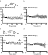
Synaptic inhibition and firing mode. a, An EPSP burst-pairing experiment is shown with picrotoxin (10 μm), which was intracellularly applied to block synaptic inhibition. Sample EPSPs are drawn from the corresponding time points indicated in the time series plot. b, Average time course of normalized EPSP amplitudes with synaptic inhibition blocked. Note the long-lasting diminution of the EPSPs, which is indicative of long-term depression. c, EPSP amplitude time series before and after a pairing episode (n = 70) with individual action potentials delayed by +10 msec. Sample EPSPs in control and after conditioning are shown at the top. d, Mean time course of all normalized EPSP amplitudes in single-spike pairing experiments. Note the long-lasting enhancement of the conditioned EPSPs, indicative of long-term potentiation. Dashed lines indicate baseline, and black horizontal bars indicate the pairing period.
Figure 3.
Figure 3.
Summary scatter plots for all experiments in which EPSPs were paired with +10 msec bursts of action potentials in normal solution (a) or with synaptic inhibition blocked (b) and in which EPSPs were paired with individual action potentials (AP) delayed by +10 msec (c). All pairings were repeated 70 times at 0.2 Hz. The diagonals represent the identity line. Data from perforated patch-clamp recordings (circles) and whole-cell recordings (triangles) are symbol coded. Note that the majority of EPSPs were depressed after the burst pairings. However, with the single-spike pairing protocol, most EPSPs were potentiated. Bar graphs show average EPSP amplitudes in control (black) and after conditioning (white). *Significantly different.
Figure 4.
Figure 4.
Burst-LTD depends on timing and number of pairings. a, The relative size (mean ± SEM) of conditioned EPSPs at different burst-pairing intervals. • p < 0.05, significantly depressed. b, Summary bar graph for relative EPSP depression (in %) for different numbers of pairings as indicated. * p < 0.05, significantly depressed.
Figure 5.
Figure 5.
Burst-LTD is associative. a, Sample traces and amplitude time series of an experiment in which EPSPs were repetitively evoked at 0.2 Hz while the postsynaptic cell remained silent. b, Average time series of all unconditioned EPSP amplitudes normalized to control. Note the lack of a significant change in EPSP size. c, Sample traces and amplitude time series of EPSPs for a conditioning protocol during which synaptic stimulation was interrupted while the postsynaptic neuron was activated. d, Average time course of normalized EPSP amplitudes from different cells. Note the lack of a systematic change in EPSP size. Dashed lines indicate baseline, and horizontal black bars indicate the pairing period.
Figure 6.
Figure 6.
Burst-LTD of potentiated synapses. a, Amplitude time series and sample EPSPs before and after conditioning with a +10 msec single action potential followed by pairings with a +10 msec delayed burst. b, Average time course of EPSPs normalized to control in different cells. Note the pairing-induced potentiation that is followed by depression after conditioning with a burst. Dashed lines indicate baseline, and horizontal black bars indicate the pairing period.
Figure 7.
Figure 7.
Burst-LTD requires activation of mGlu receptors. a, Amplitude time series and sample traces of evoked EPSPs in control and after 70 burst pairings in the presence of the NMDA receptor antagonist APV (50 μm). b, Average time course of normalized EPSPs in different cells with APV. Note the significant EPSP depression despite the presence of the NMDA receptor antagonist. c, Experiments similar to those in a but in the presence of the metabotropic gluta-mate receptor antagonist MCPG (0.25 mm). d, The amplitude time series of normalized EPSPs reveals the lack of a systematic change of EPSPs after blocking metabotropic glutamate receptors. Dashed lines indicate baseline, and black horizontal bars indicate the pairing period.
Figure 8.
Figure 8.
Burst-LTD requires activation of Ca2+ and Na+ channels. a, b, EPSP-burst pairings in the presence of the T-type calcium channel antagonist Ni2+ (0.1mm). a, EPSP amplitude time series and sample EPSPs are shown for an individual experiment. b, Average time course of all experiments with EPSP amplitudes normalized to control. Note that with T-channels blocked, EPSPs were not significantly altered after the pairings. c, d, Similar experiment as above but with the sodium channel blocker QX-314 (10 mm) added to the pipette solution. Note again the lack of a systematic change in EPSP size as shown for an individual cell (c) and for the entire population (d). Dashed lines indicate baseline, and horizontal black bars indicate the pairing period.

References

    1. Abbott LF, Nelson SB (2000) Synaptic plasticity: taming the beast. Nat Neurosci 3: 1178-1183. - PubMed
    1. Akaike N, Hattori K, Oomura Y, Carpenter DO (1985) Bicuculline and picrotoxin block gamma-aminobutyric acid-gated Cl- conductance by different mechanisms. Experientia 41: 70-71. - PubMed
    1. Bear MF, Press WA, Connors BW (1992) Long-term potentiation in slices of kitten visual cortex and the effects of NMDA receptor blockade. J Neurophysiol 67: 841-851. - PubMed
    1. Braunewell KH, Manahan-Vaughan D (2001) Long-term depression: a cellular basis for learning? Rev Neurosci 12: 121-140. - PubMed
    1. Connors BW, Gutnick MJ, Prince DA (1982) Electrophysiological properties of neocortical neurons in vitro. J Neurophysiol 48: 1302-1320. - PubMed

Publication types

MeSH terms

Substances