

An experiment of nature: brain anatomy parallels cognition and behavior in Williams syndrome
- PMID: 15163693
- PMCID: PMC3061615
- DOI: 10.1523/JNEUROSCI.5272-03.2004
An experiment of nature: brain anatomy parallels cognition and behavior in Williams syndrome
Abstract
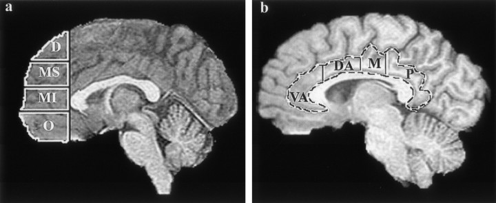
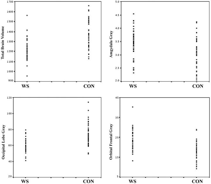
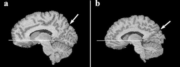
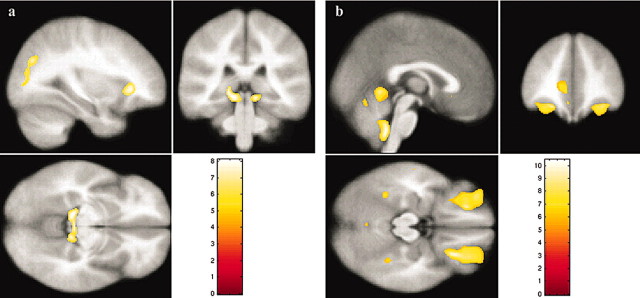
Williams syndrome (WS) is a neurogenetic-neurodevelopmental disorder characterized by a highly variable and enigmatic profile of cognitive and behavioral features. Relative to overall intellect, affected individuals demonstrate disproportionately severe visual-spatial deficits and enhanced emotionality and face processing. In this study, high-resolution magnetic resonance imaging data were collected from 43 individuals with WS and 40 age- and gender-matched healthy controls. Given the distinct cognitive-behavioral dissociations associated with this disorder, we hypothesized that neuroanatomical integrity in WS would be diminished most in regions comprising the visual-spatial system and most "preserved" or even augmented in regions involved in emotion and face processing. Both volumetric analysis and voxel-based morphometry were used to provide convergent approaches for detecting the hypothesized WS neuroanatomical profile. After adjusting for overall brain volume, participants with WS showed reduced thalamic and occipital lobe gray matter volumes and reduced gray matter density in subcortical and cortical regions comprising the human visual-spatial system compared with controls. The WS group also showed disproportionate increases in volume and gray matter density in several areas known to participate in emotion and face processing, including the amygdala, orbital and medial prefrontal cortices, anterior cingulate, insular cortex, and superior temporal gyrus. These findings point to specific neuroanatomical correlates for the unique topography of cognitive and behavioral features associated with this disorder.
Figures
References
-
- Adolphs R (2003) Is the human amygdala specialized for processing social information? Ann NY Acad Sci 985: 326-340. - PubMed
-
- Atkinson J, King J, Braddick O, Nokes L, Anker S, Braddick F (1997) A specific deficit of dorsal stream function in Williams' syndrome. NeuroReport 8: 1919-1922. - PubMed
-
- Atkinson J, Anker S, Braddick O, Nokes L, Mason A, Braddick F (2001) Visual and visuospatial development in young children with Williams syndrome. Dev Med Child Neurol 43: 330-337. - PubMed
-
- Bellugi U, Lichtenberger L, Mills D, Galaburda A, Korenberg JR (1999) Bridging cognition, the brain and molecular genetics: evidence from Williams syndrome. Trends Neurosci 22: 197-207. - PubMed
Publication types
MeSH terms
Grants and funding
LinkOut - more resources
Full Text Sources