. 2004 Jun;78(12):6304-12.
doi: 10.1128/JVI.78.12.6304-6312.2004.

PB1-F2, an influenza A virus-encoded proapoptotic mitochondrial protein, creates variably sized pores in planar lipid membranes

Affiliations

PB1-F2, an influenza A virus-encoded proapoptotic mitochondrial protein, creates variably sized pores in planar lipid membranes

A N Chanturiya et al. J Virol. 2004 Jun.

Abstract

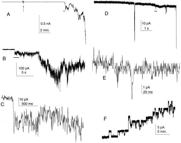
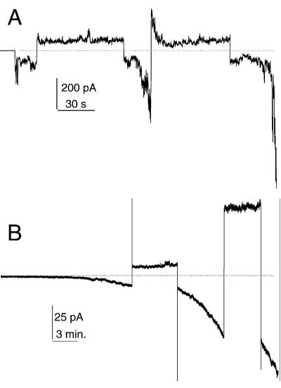
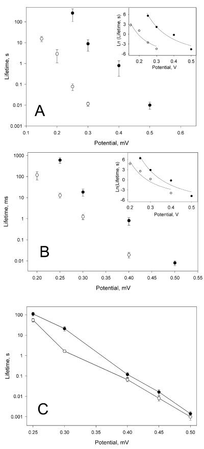
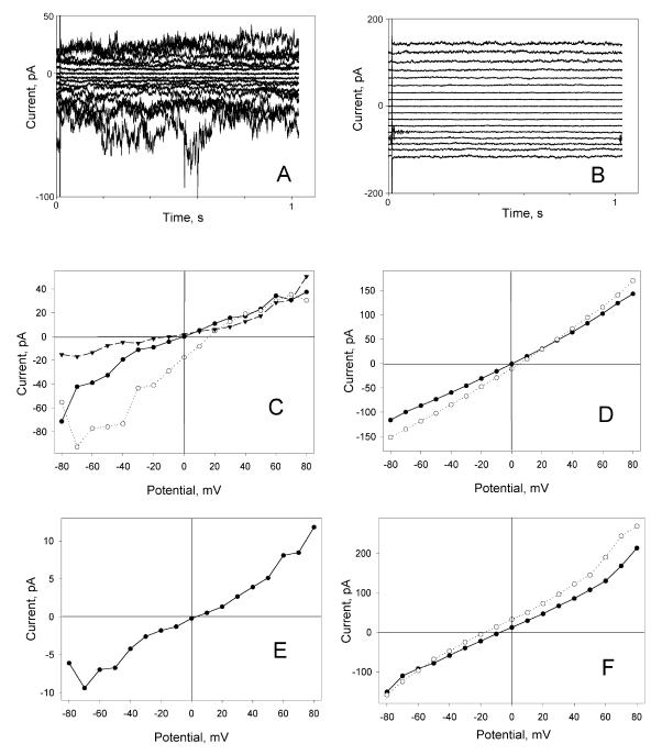
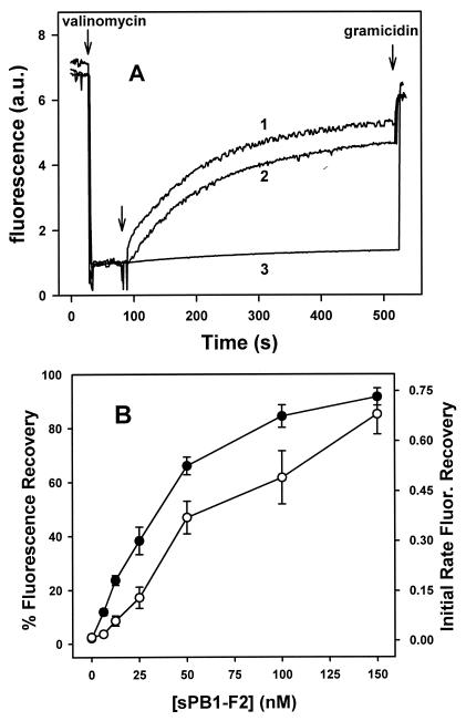
A frameshifted region of the influenza A virus PB1 gene encodes a novel protein, termed PB1-F2, a mitochondrial protein that can induce cell death. Many proapoptotic proteins are believed to act at the mitochondrial outer membrane to form an apoptotic pore with lipids. We studied the interaction of isolated, synthetic PB1-F2 (sPB1-F2) peptide with planar phospholipid bilayer membranes. The presence of nanomolar concentrations of peptide in the bathing solution induced a transmembrane conductance that increased in a potential-dependent manner. Positive potential on the side of protein addition resulted in a severalfold increase in the rate of change of membrane conductance. sPB1-F2-treated membranes became permeable to monovalent cations, chloride, and to a lesser extent, divalent ions. Despite various experimental conditions, we did not detect the distinctive conductance levels typical of large, stable pores, protein channels, or even pores that are partially proteinaceous. Rather, membrane conductance induced by sPB1-F2 fluctuated and visited almost all conductance values. sPB1-F2 also dramatically decreased bilayer stability in an electric field, consistent with a decrease in the line tension of a lipidic pore. Since similar membrane-destabilizing profiles are seen with proapoptotic proteins (e.g., Bax) and the cytoplasmic helix of human immunodeficiency virus gp41, we suggest that the basis for sPB1-F2-induced cell death may be the permeabilization and destabilization of mitochondrial membranes, leading to macromolecular leakage and apoptosis.


Figures

FIG. 1.
FIG. 1.
Increase in conductance induced by sPB1-F2 in membrane. (A) Low-resolution record of the experiment. The arrow indicates the addition of 20 nM (final concentration) sPB1-F2 to the cis side of the membrane. (B to E) Medium-resolution (B and D) and high-resolution (C and E) records of sPB1-F2-induced fluctuations in conductance. Horizontal black bars in panels A, B, and D show the approximate position of the fragment expanded in the next panel. Discrete conductance levels visible in panel E are due to finite digitizer resolution. Recordings shown in panels A to C are obtained on a 0.2-mm-diameter membrane formed from DPhC in a solution consisting of 150 mM KCl, 1 mM MgCl2, 1 mM EGTA, 1 mM dithiothreitol, and 20 mM HEPES (pH 7.4). Recordings shown in panels D and E are from DOPE-DOPC membrane on a 5-μm-diameter glass micropipette tip with 100 mM KCl-10 mM HEPES (pH 7.5) inside and outside the pipette. The membrane potential in both cases was −50 mV. (F) Membrane permeabilization by S. aureus alpha-toxin. The final concentration of S. aureus alpha-toxin was 30 nM. The membrane potential was +50 mV. The DOPC-DOPE membrane was formed in 0.1 M KCl-5 mM HEPES (pH 7.5). For all experiments, protein was added to the grounded compartments.
FIG. 2.
FIG. 2.
Potential dependence of sPB1-F2 insertion into membrane. (A) sPB1-F2 (10 nM) was added to the cis (ground) side of the membrane approximately 5 min before the beginning of conductance increase. The membrane was formed from DPhC. (While it was not possible to obtain long records with DOPC-DOPE [50%/50%, wt/wt] membrane due to lower stability, qualitatively, the results were similar.) (B) sPB1-F2 (80 nM) was added approximately 15 and 30 min before the beginning of the record. The membrane was formed from DOPC-DOPE-DOPS (25%/25%/50% [wt/wt/wt]). In both cases, the membrane bathing solution contains 100 mM KCl-10 mM HEPES (pH 7.4). The horizontal line indicates zero current. The membrane potential was switched several times (first, −50 and second, +50 mV) and kept constant during positive or negative fragments of current traces.
FIG. 3.
FIG. 3.
Potential dependence of protein-induced decrease in membrane lifetime. (A) Effect of sPB1-F2 on membrane formed from neutral lipid mixture DOPE-DOPC (50%/50% [wt/wt]). (B) Effect of sPB1-F2 on membrane formed from the mixture that contains charged lipid DOPE-DOPC-DOPS (25%/25%/50% [wt/wt/wt]). (C) Effect of S. aureus alpha-toxin on membrane formed from egg phosphatidylcholine-cholesterol (3/1). The mean values ± standard errors (error bars) from 10 to 15 experiments are plotted in conventional form. The inserts show the same data and theoretical fit, plotted as Ln(lifetime) versus voltage in order to balance curve-fitting error over the wide range of experimental values. Control experiments (•) and experiments performed with 10 nM sPB1-F2 (○) (A and B) or 12 nM alpha-toxin (C) on both sides of membrane are shown. The membrane bathing solution in panels A and B was 0.1 M KCl-10 mM HEPES (pH 7.4); the membrane bathing solution in panel C was 0.1 M KCl-1 mM EDTA-10 mM Tris citrate (pH 7.5).
FIG. 4.
FIG. 4.
Current voltage characteristics of sPB1-F2-modified membranes. (A and B) Current traces of sPB1-F2-treated membrane at −80 to +80 mV potentials (cis ground) with 10 mV increment. The membrane was DPhC membrane bathed in symmetrical 0.1 M NaCl solution (A) or DOPE-DOPC-DOPS (25%/25%/50% [wt/wt/wt]) membrane bathed in symmetrical 0.1 M KCl solution (B). (C) Dependence of electric current (I) and electrical potential (V) of sPB1-F2-treated DPhC membrane in symmetrical 0.1 M NaCl (•), 0.1 M NaCl in the trans compartment and 0.35 M NaCl in the cis compartment (○), and after the addition of 5 mM CdCl2 to the cis compartment ▾. (D) Dependence of I and V of sPB1-F2-treated DOPE-DOPC-DOPS (25%/25%/50% [wt/wt/wt]) membrane in symmetrical 0.1 M KCl (•) and 0.1 M KCl in the trans compartment and 0.25 M KCl in the cis compartment (○). (E) Dependence of I and V on sPB1-F2-treated DPhC membrane in 0.1 M NaCl in the cis compartment and 0.1 M KCl in the trans compartment. (F) Dependence of I and V of sPB1-F2-treated DPhC membrane formed in asymmetrical solutions, 5 mM MgCl2 in the cis compartment and 25 mM MgCl2 in the trans compartment (○) and after the addition of 5 mM CdCl2 to the cis compartment (•).
FIG. 5.
FIG. 5.
Loss of transmembrane potential induced by PB1-F2 in LUV. (A) DOPC-cholesterol (3/1) LUV with fluorescent dye diSC3-(5) were prepared as described in Materials and Methods. The addition of valinomycin (first arrow) created a negative-inside transmembrane potential (Δψ), which led to quenching of the dye's fluorescence. At the time indicated by the second arrow, 50 nM S. aureus alpha-toxin (curve 1), 50 nM sPB1-F2 (curve 2), or buffer alone (curve 3) was added, and Δψ loss was monitored by recovery of the fluorescence signal. Finally, gramicidin was added (third arrow) to completely dissipate the LUV transmembrane potential. The similarity in the time courses of potential dissipation by PB1-F2 and alpha-toxin indicates that it is more likely that sPB1-F2 forms pores than ruptures membrane. Fluorescence is given in arbitrary units (a.u.). (B) Dissipation of vesicular Δψ as a function of sPB1-F2 concentration. DOPC LUV were treated with different amounts of sPB1-F2, and the values of the maximal extent (•) and initial rate (○) of diSC3-(5) fluorescence (Fluor.) recovery were estimated as explained in Materials and Methods. Data are averages ± standard errors (error bars) of triplicate determinations of at least two independent LUV populations. Other conditions are as explained above for panel A. Note that the extravesicular compartment is equivalent to cis or the ground side of the planar membrane.

References

    1. Abidor, I. G., V. B. Arakelyan, L. V. Chernomordik, Y. A. Chizmadzhev, V. F. Pastushenko, and M. R. Tarasevich. 1979. Electrical breakdown of BLM: main experimental facts and their qualitative discussion. Bioelectrochem. Bioenerget. 6:37-52.
    1. Barlett, G. R. 1959. Phosphorus assay in column chromatography. J. Biol. Chem. 234:466-468. - PubMed
    1. Basañez, G., A. Nechushtan, O. Drozhinin, A. Chanturiya, E. Choe, S. Tutt, K. A. Wood, Y.-T. Hsu, J. Zimmerberg, and R. J. Youle. 1999. Bax, but not Bcl-xL, decreases the lifetime of planar phospholipid bilayer membranes at subnanomolar concentrations. Proc. Natl. Acad. Sci. USA 96:5492-5497. - PMC - PubMed
    1. Basañez, G., J. C. Sharpe, J. Galanis, T. B. Brandt, J. M. Hardwick, and J. Zimmerberg. 2002. Bax-type apoptotic proteins porate pure lipid bilayers through a mechanism sensitive to intrinsic monolayer curvature. J. Biol. Chem. 277:49360-49365. - PubMed
    1. Basañez, G., J. Zhang, B. N. Chau, G. I. Maksaev, V. A. Frolov, T. A. Brandt, J. Burch, J. M. Hardwick, and J. Zimmerberg. 2001. Pro-apoptotic cleavage products of Bcl-xL form cytochrome c-conducting pores in pure lipid membranes. J. Biol. Chem. 276:31083-31091. - PubMed

Publication types

MeSH terms