. 2004 Jun;78(12):6370-80.
doi: 10.1128/JVI.78.12.6370-6380.2004.

Intramembrane proteolysis and endoplasmic reticulum retention of hepatitis C virus core protein

Affiliations

Intramembrane proteolysis and endoplasmic reticulum retention of hepatitis C virus core protein

Kiyoko Okamoto et al. J Virol. 2004 Jun.

Abstract

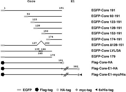
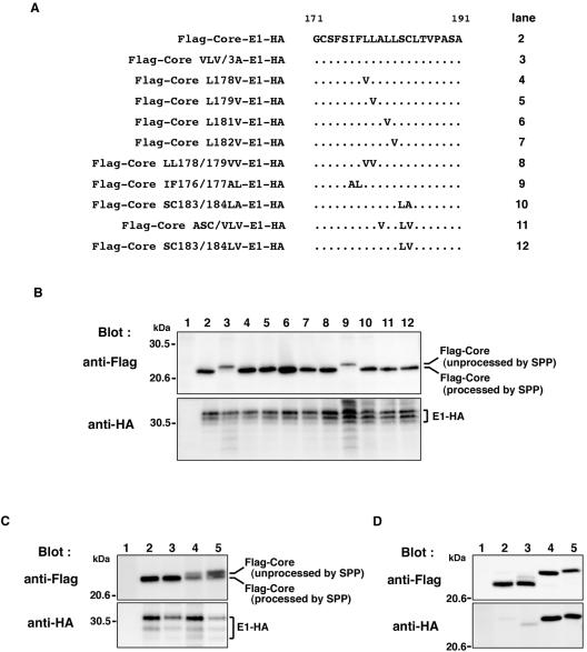
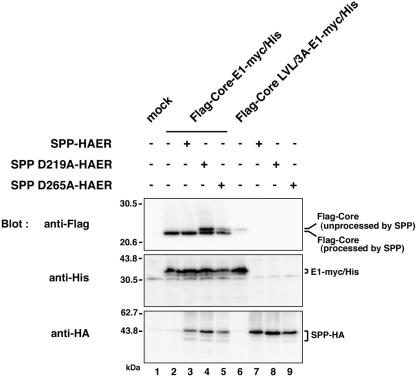
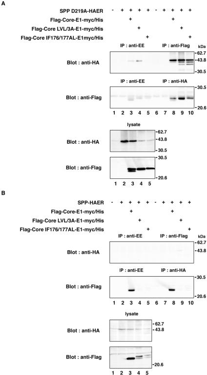
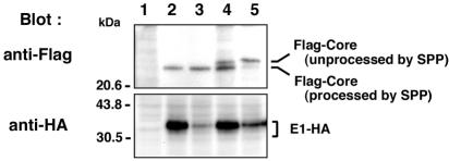
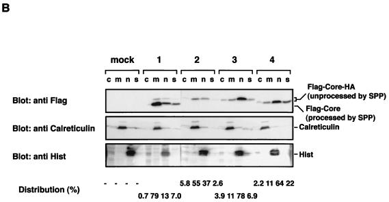
Hepatitis C virus (HCV) core protein is suggested to localize to the endoplasmic reticulum (ER) through a C-terminal hydrophobic region that acts as a membrane anchor for core protein and as a signal sequence for E1 protein. The signal sequence of core protein is further processed by signal peptide peptidase (SPP). We examined the regions of core protein responsible for ER retention and processing by SPP. Analysis of the intracellular localization of deletion mutants of HCV core protein revealed that not only the C-terminal signal-anchor sequence but also an upstream hydrophobic region from amino acid 128 to 151 is required for ER retention of core protein. Precise mutation analyses indicated that replacement of Leu(139), Val(140), and Leu(144) of core protein by Ala inhibited processing by SPP, but cleavage at the core-E1 junction by signal peptidase was maintained. Additionally, the processed E1 protein was translocated into the ER and glycosylated with high-mannose oligosaccharides. Core protein derived from the mutants was translocated into the nucleus in spite of the presence of the unprocessed C-terminal signal-anchor sequence. Although the direct association of core protein with a wild-type SPP was not observed, expression of a loss-of-function SPP mutant inhibited cleavage of the signal sequence by SPP and coimmunoprecipitation with unprocessed core protein. These results indicate that Leu(139), Val(140), and Leu(144) in core protein play crucial roles in the ER retention and SPP cleavage of HCV core protein.


Figures

FIG. 1.
FIG. 1.
Expression plasmids used in this study. The genes encoding HCV proteins and their mutants were cloned into pcDNA3.1FlagHA, pcDNA3.1/myc-His C, or pEGFP-C3 as described in Materials and Methods. Other plasmids are described in the text or in the other figure legends.
FIG. 2.
FIG. 2.
Intracellular localization of EGFP-Core mutants. EGFP-Core and its deletion mutants were coexpressed with ER-DsRed in HeLa cells, and the localization of core proteins was examined by confocal microscopy.
FIG. 3.
FIG. 3.
Identification of the region responsible for processing of the signal sequence of HCV core protein by SPP and signal peptidase. (A) The hydrophobicity profile of HCV core protein was predicted by the method of Kyte and Doolittle (20). Hope and McLauchlan separated the HCV core protein into three regions, domains 1 to 3 (12). Two hydrophobic regions are predicted in the regions from amino acid 128 to 151 and from amino acid 164 to 186 in the C terminus of the HCV core protein. Mutations and deletions in the region from amino acid 128 to 151 of Flag-Core-HA and Flag-Core-E1-HA constructs are indicated. Dots indicate unchanged amino acids. (B) Expression of Flag-Core-HA polyproteins with changes of Ala191 to Arg in 293T cells. Flag-Core-HA (lanes 2 and 11), Flag-Core L139A-HA (lane 3), Flag-Core V140A-HA (lane 4), Flag-Core L144A-HA (lane 5), Flag-Core LV139/140AA-HA (lane 6), Flag-Core LL139/144AA-HA (lane 7), Flag-Core VL140/144AA-HA (lane 8), Flag-Core LVL/3A-HA (lane 9), and Flag-Core Δ128-151-HA (lane 10) were analyzed by immunoblotting with anti-Flag (upper panel) or anti-HA (lower panel) antibody. Cells transfected with an empty plasmid were used as a negative control (lane 1). (C) Expression of Flag-Core-E1-HA mutants in 293T cells. Flag-Core-E1-HA (lanes 2 and 11), Flag-Core L139A-E1-HA (lane 3), Flag-Core V140A-E1-HA (lane 4), Flag-Core L144A-E1-HA (lane 5), Flag-Core LV139/140AA-E1-HA (lane 6), Flag-Core LL139/144AA-E1-HA (lane 7), Flag-Core VL140/144AA-E1-HA (lane 8), Flag-Core LVL/3A-E1-HA (lane 9), and Flag-Core Δ128-151-E1-HA (lane 10) were analyzed by immunoblotting with anti-Flag (upper panel) or anti-HA (lower panel) antibody. The asterisk indicates unprocessed Flag-Core Δ128-151. Cells transfected with an empty plasmid were used as a negative control (lane 1). (D) The deglycosylation procedure is described in Materials and Methods. After transfection, cell lysates were immunoprecipitated with anti-HA antibody and immunoprecipitates were digested with Endo H (upper panel) or PNGase F (lower panel). Following digestion, proteins were separated by SDS-polyacrylamide gel electrophoresis, and material from cells transfected with vector (lane 1), Flag-Core-E1-HA (lane 2), Flag-Core LVL/3A-E1-HA (lane 3), and Flag-Core Δ128-151-E1-HA (lane 4) was detected by blotting with anti-HA. Nontreated and Endo H- or PNGase F-treated samples are indicated by − and +, respectively. Asterisks indicate mouse IgG heavy chains.
FIG. 4.
FIG. 4.
Amino acid residues essential for SPP cleavage of the HCV core protein signal sequence of genotype 1a and 1b strains. (A) Mutations in the amino acid residues in the signal sequence of Flag-Core-E1-HA are indicated. Dots indicate unchanged amino acids. (B) Flag-Core-E1-HA (lane 2), Flag-Core LVL/3A-E1-HA (lane 3), Flag-Core L178V-E1-HA (lane 4), Flag-Core L179V-E1-HA (lane 5), Flag-Core L181V-E1-HA (lane 6), Flag-Core L182V-E1-HA (lane 7), Flag-Core LL178/179VV-E1-HA (lane 8), Flag-Core IF176/177AL-E1-HA (lane 9), Flag-Core SC183/184LA-E1-HA (lane 10), Flag-Core ASC/VLV-E1-HA (lane 11), or Flag-Core SC183/184LV-E1-HA (lane 12) was expressed in 293T cells. (C) The gene encoding core and E1 polyprotein of the genotype 1a H77c strain of HCV was introduced into pcDNA3.1FlagHA. Flag-H77c Core-E1-HA (lane 2), Flag-H77c Core ASC/VLV-E1-HA (lane 3), Flag-H77c Core LVL/3A-E1-HA (lane 4), or Flag-H77c Core IF176/177AL-E1-HA (lane 5) was expressed in BHK cells. Cell lysates were analyzed by immunoblotting with anti-Flag (upper panel) and anti-HA (lower panel) antibodies. (D) Expression of Flag-Core 191-HA mutants in 293T cells. The gene encoding core protein with a change of Ala191 to Arg was introduced into pcDNA3.1FlagHA. Flag-Core-HA (lane 2), Flag-Core ASC/VLV-HA (lane 3), Flag-Core LVL/3A-HA (lane 4), and Flag-Core IF176/177AL-HA (lane 5) were analyzed by immunoblotting with anti-Flag (upper panel) and anti-HA (lower panel) antibodies. Cells transfected with an empty plasmid were used as a negative control (lanes 1 in panels B, C, and D).
FIG. 5.
FIG. 5.
Effect of loss-of-function mutants of SPP on the processing of the signal sequence of HCV core protein. SPP-HAER, SPPD219A-HAER, or SPPD265A-HAER was coexpressed with Flag-Core-E1-myc/His or Flag-Core LVL/3A-E1-myc/His in 293T cells. Cell lysates were analyzed by immunoblotting with anti-Flag (upper panel), anti-His6 (middle panel), or anti-HA (lower panel) antibody. + and −, presence or absence of each plasmid, respectively. Lane 1, mock; lanes 2, 6, 7, 8, and 9, single expression of Flag-Core-E1-myc/His, Flag-Core LVL/3A-E1-myc/His, SPP-HAER, SPPD219A-HAER, and SPPD265A-HAER, respectively; lanes 3 to 5, coexpression of Flag-Core-E1-myc/His with SPP-HAER, SPPD219A-HAER, and SPPD265A-HAER, respectively.
FIG. 6.
FIG. 6.
Interaction of HCV core protein with SPP. SPP D219A-HAER (A) or SPP-HAER (B) was coexpressed with Flag-Core-E1-myc/His, Flag-Core LVL/3A-E1-myc/His, or Flag-Core IF176/177AL-E1-myc/His in 293T cells and immunoprecipitated (IP) with anti-Flag or anti-HA antibody. The immunoprecipitates were analyzed by immunoblotting with anti-HA or anti-Flag antibody. As a control, immunoprecipitation was carried out with anti-EE antibody. + and −, presence or absence of each plasmid, respectively.
FIG. 7.
FIG. 7.
Processing of HCV core-E1 polyprotein in the human hepatoma cell line FLC-4. Flag-Core-E1-HA (lane 2), Flag-Core ASC/VLV-E1-HA (lane 3), Flag-Core LVL/3A-E1-HA (lane 4), or Flag-Core IF176/177AL-E1-HA (lane 5) was expressed in FLC-4 cells and analyzed by immunoblotting with anti-Flag (upper panel) or anti-HA (lower panel) antibody. Cells transfected with an empty plasmid were used as a negative control (lane 1).
FIG. 8.
FIG. 8.
Localization of mutant HCV core proteins. (A) Putative processing mechanisms of wild-type and mutant core proteins is illustrated at the top. EGFP-Core-191 (column 1), EGFP-Core IF176/177AL (column 2), EGFP-Core LVL/3A (column 3), and EGFP-Core Δ128-151 (column 4) were coexpressed with ER-DsRed in HeLa cells, and subcellular localization of core proteins was examined by confocal microscopy. (B) Subcellular fractionation of HeLa cells transfected with plasmids encoding Flag-Core-HA polyproteins. Cells transfected with an empty plasmid (lane M) or plasmid encoding Flag-Core 191-HA (lanes 1), Flag-Core IF176/177AL-HA (lanes 2), Flag-Core LVL/3A-HA (lanes 3), or Flag-Core Δ128-151-HA (lanes 4) were extracted into four fractions, as described in Materials and Methods. Each fraction was concentrated and subjected to immunoblotting with anti-Flag antibody (upper panel). Lanes c, m, n and s, cytosol, membrane-organelle, nuclear, and cytoskeleton fractions, respectively. Calreticulin and histone (His) were used as markers for membrane-organelle and nuclear fractions, respectively. To determine the distributed ratio of processed and unprocessed core proteins in each fraction, the density of core protein in each fraction was measured and is indicated as a percentage at each bottom of lane.
FIG. 8.
FIG. 8.
Localization of mutant HCV core proteins. (A) Putative processing mechanisms of wild-type and mutant core proteins is illustrated at the top. EGFP-Core-191 (column 1), EGFP-Core IF176/177AL (column 2), EGFP-Core LVL/3A (column 3), and EGFP-Core Δ128-151 (column 4) were coexpressed with ER-DsRed in HeLa cells, and subcellular localization of core proteins was examined by confocal microscopy. (B) Subcellular fractionation of HeLa cells transfected with plasmids encoding Flag-Core-HA polyproteins. Cells transfected with an empty plasmid (lane M) or plasmid encoding Flag-Core 191-HA (lanes 1), Flag-Core IF176/177AL-HA (lanes 2), Flag-Core LVL/3A-HA (lanes 3), or Flag-Core Δ128-151-HA (lanes 4) were extracted into four fractions, as described in Materials and Methods. Each fraction was concentrated and subjected to immunoblotting with anti-Flag antibody (upper panel). Lanes c, m, n and s, cytosol, membrane-organelle, nuclear, and cytoskeleton fractions, respectively. Calreticulin and histone (His) were used as markers for membrane-organelle and nuclear fractions, respectively. To determine the distributed ratio of processed and unprocessed core proteins in each fraction, the density of core protein in each fraction was measured and is indicated as a percentage at each bottom of lane.

References

    1. Aizaki, H., Y. Aoki, T. Harada, K. Ishii, T. Suzuki, S. Nagamori, G. Toda, Y. Matsuura, and T. Miyamura. 1998. Full-length complementary DNA of hepatitis C virus genome from an infectious blood sample. Hepatology 27:621-627. - PubMed
    1. Bukh, J., R. H. Purcell, and R. H. Miller. 1994. Sequence analysis of the core gene of 14 hepatitis C virus genotypes. Proc. Natl. Acad. Sci. USA 91:8239-8243. - PMC - PubMed
    1. Chang, S. C., J. H. Yen, H. Y. Kang, M. H. Jang, and M. F. Chang. 1994. Nuclear localization signals in the core protein of hepatitis C virus. Biochem. Biophys. Res. Commun. 205:1284-1290. - PubMed
    1. Chen, C. M., L. R. You, L. H. Hwang, and Y. H. Lee. 1997. Direct interaction of hepatitis C virus core protein with the cellular lymphotoxin-beta receptor modulates the signal pathway of the lymphotoxin-beta receptor. J. Virol. 71:9417-9426. - PMC - PubMed
    1. Choo, Q. L., G. Kuo, A. J. Weiner, L. R. Overby, D. W. Bradley, and M. Houghton. 1989. Isolation of a cDNA clone derived from a blood-borne non-A, non-B viral hepatitis genome. Science 244:359-362. - PubMed

Publication types

Substances

LinkOut - more resources