. 2004 Jun;78(12):6469-79.
doi: 10.1128/JVI.78.12.6469-6479.2004.

Inter- and intragenus structural variations in caliciviruses and their functional implications

Affiliations

Inter- and intragenus structural variations in caliciviruses and their functional implications

Rong Chen et al. J Virol. 2004 Jun.

Abstract

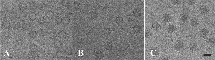
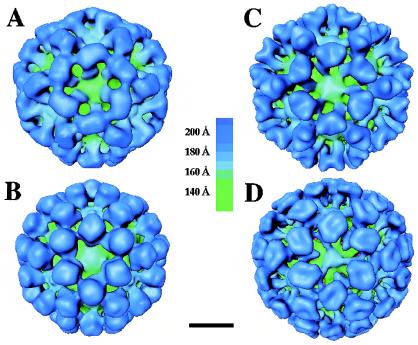
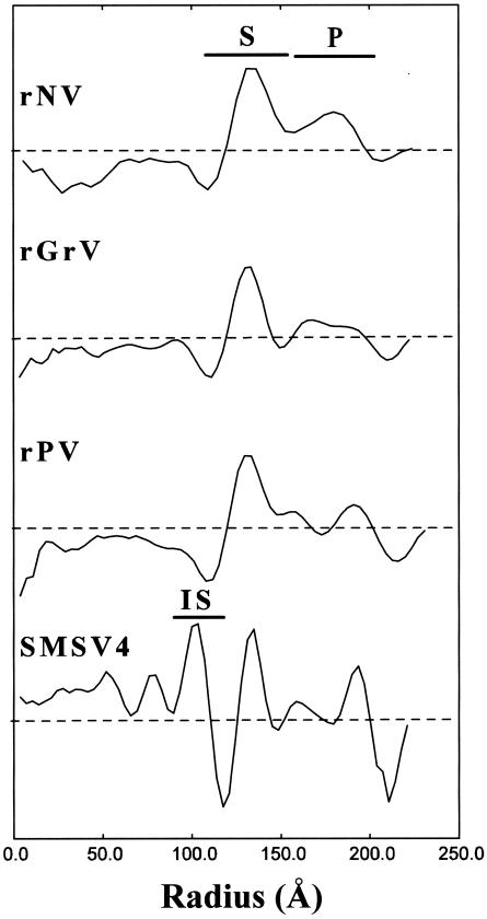
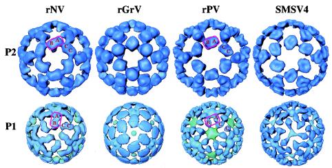
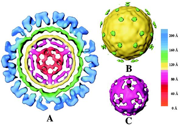
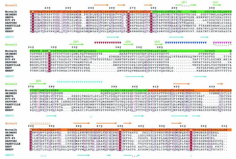
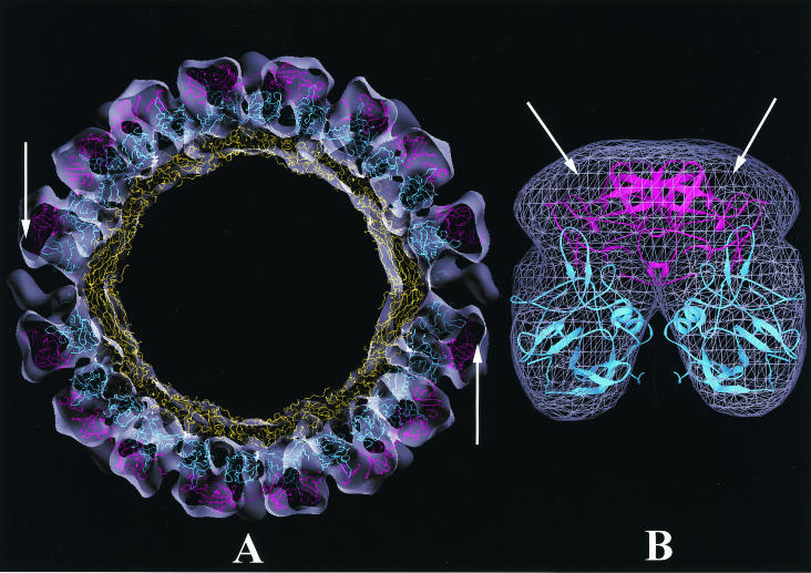
The family Caliciviridae is divided into four genera and consists of single-stranded RNA viruses with hosts ranging from humans to a wide variety of animals. Human caliciviruses are the major cause of outbreaks of acute nonbacterial gastroenteritis, whereas animal caliciviruses cause various host-dependent illnesses with a documented potential for zoonoses. To investigate inter- and intragenus structural variations and to provide a better understanding of the structural basis of host specificity and strain diversity, we performed structural studies of the recombinant capsid of Grimsby virus, the recombinant capsid of Parkville virus, and San Miguel sea lion virus serotype 4 (SMSV4), which are representative of the genera Norovirus (genogroup 2), Sapovirus, and Vesivirus, respectively. A comparative analysis of these structures was performed with that of the recombinant capsid of Norwalk virus, a prototype member of Norovirus genogroup 1. Although these capsids share a common architectural framework of 90 dimers of the capsid protein arranged on a T=3 icosahedral lattice with a modular domain organization of the subunit consisting of a shell (S) domain and a protrusion (P) domain, they exhibit distinct differences. The distally located P2 subdomain of P shows the most prominent differences both in shape and in size, in accordance with the observed sequence variability. Another major difference is in the relative orientation between the S and P domains, particularly between those of noroviruses and other caliciviruses. Despite being a human pathogen, the Parkville virus capsid shows more structural similarity to SMSV4, an animal calicivirus, suggesting a closer relationship between sapoviruses and animal caliciviruses. These comparative structural studies of caliciviruses provide a functional rationale for the unique modular domain organization of the capsid protein with an embedded flexibility reminiscent of an antibody structure. The highly conserved S domain functions to provide an icosahedral scaffold; the hypervariable P2 subdomain may function as a replaceable module to confer host specificity and strain diversity; and the P1 subdomain, located between S and P2, provides additional fine-tuning to position the P2 subdomain.


Figures

FIG. 1.
FIG. 1.
Cryo-EM images of rGrV (A), rPV (B), and SMSV4 (C) particles embedded in vitreous ice. SMSV4 virions are substantially darker in the middle than the recombinant capsids (A and B) due to the presence of the RNA genome. Bar = 400 Å.
FIG. 2.
FIG. 2.
Surface representations of the structures of rNV (A), rGrV (B), rPV (C), and SMSV4 (D) at a resolution of ∼22 Å viewed along the icosahedral threefold axis. Structures are radially color coded according to the chart shown in the middle. Bar = 100 Å.
FIG. 3.
FIG. 3.
Radial density plots of rNV, rGrV, rPV, and SMSV4 computed from their respective cryo-EM density maps. The dashed horizontal lines represent an average radial density of zero. The radial locations of the S and P domains as well as the IS are denoted.
FIG. 4.
FIG. 4.
Structural comparisons of the P1 and P2 subdomains among different caliciviruses. All of the surface representations are radially colored and viewed in the same orientation as in Fig. 2. The locations of the A, B, and C subunits are denoted. In the structures of rNV and rGrV, the P1 subdomain participates in intradimeric interactions, whereas in the structures of rPV and SMSV4, the same region is involved in interdimeric interactions. In rNV and rGrV, the P domain, shown inside a rectangular box, undergoes a twist of about 45° (compare the upper and lower panels), whereas in rPV and SMSV4, such a twist is not present.
FIG. 5.
FIG. 5.
Internal features in the SMSV4 virion structure. (A) Central equatorial section (20 Å in thickness) from SMSV4 map radially color coded according to the chart shown on the right. The P and S domain densities are shown in blue and green, respectively; the IS beneath the S-domain shell is shown in yellow. (B) Surface representation of IS and the weak densities that connect the IS with the S domain. (C) SMSV4 density map between the radii of 40 and 85 Å.
FIG. 6.
FIG. 6.
Sequence alignment in the P domain, derived by comparing 30 calicivirus capsid protein sequences with the program CLUSTALW. Only eight calicivirus sequences (two from each genus), including those of NV, SMSV4, GrV, and PV, are shown. The secondary structural elements inferred from the rNV X-ray structure are shown above the sequences. As an example, the predicted secondary structure of SMSV4 is shown below. The numbers above the sequences correspond to the residue numbers in the NV sequence. Residues which are strictly conserved and moderately conserved are shown in red and pink, respectively. The regions corresponding to the P1 and P2 subdomains are highlighted in orange and green, respectively. Possible regions of insertion in SMSV4 are shown by inverted triangles.
FIG. 7.
FIG. 7.
Fitting of rNV capsid protein coordinates into the cryo-EM density map of SMSV4. (A) Central section viewed along the icosahedral twofold axis, with the rNV capsid structure fitted into the cryo-EM density map of SMSV4 shown as a semitransparent surface. The S, P1, and P2 domains of rNV, colored in yellow, cyan, and magenta, respectively, were each treated as a rigid body during fitting and refinement. For clarity, only the Cα backbone is shown. A reasonably good fitting is seen in the S and P1 regions. The arrows indicate some extra densities in the P2 region that are not occupied by the rNV coordinates. (B) Cryo-EM densities (wired cage) of the P regions of the SMSV4 CC dimer fitted with rNV coordinates shown in a ribbon representation following the same color scheme as for panel A. Extra densities in the P2 region are indicated by arrows.

References

    1. Anonymous. 1994. The CCP4 suite: programs for protein crystallography. Acta Crystallogr. D 50:760-763. - PubMed
    1. Belliot, G., J. S. Noel, J. F. Li, Y. Seto, C. D. Humphrey, T. Ando, R. I. Glass, and S. S. Monroe. 2001. Characterization of capsid genes, expressed in the baculovirus system, of three new genetically distinct strains of “Norwalk-like viruses.” J. Clin. Microbiol. 39:4288-4295. - PMC - PubMed
    1. Bertolotti-Ciarlet, A., L. J. White, R. Chen, B. V. Prasad, and M. K. Estes. 2002. Structural requirements for the assembly of Norwalk virus-like particles. J. Virol. 76:4044-4055. - PMC - PubMed
    1. Brünger, A. T. 1993. XPLOR manual,version 3.1. Yale University, New Haven, Conn.
    1. Carter, M. J., I. D. Milton, P. C. Turner, J. Meanger, M. Bennett, and R. M. Gaskell. 1992. Identification and sequence determination of the capsid protein gene of feline calicivirus. Arch. Virol. 122:223-235. - PMC - PubMed

Publication types

MeSH terms

LinkOut - more resources