. 2004 Jun;78(12):6543-55.
doi: 10.1128/JVI.78.12.6543-6555.2004.

Lyssavirus matrix protein induces apoptosis by a TRAIL-dependent mechanism involving caspase-8 activation

Affiliations

Lyssavirus matrix protein induces apoptosis by a TRAIL-dependent mechanism involving caspase-8 activation

Raïd Kassis et al. J Virol. 2004 Jun.

Abstract

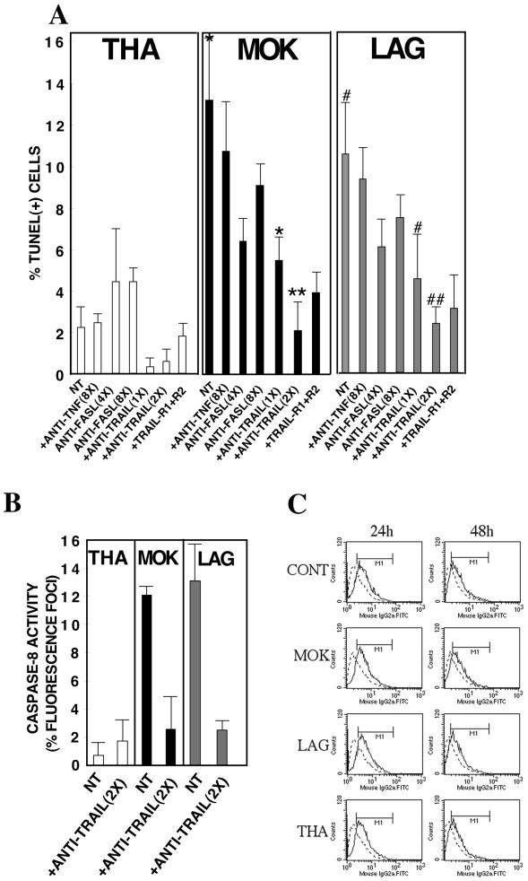
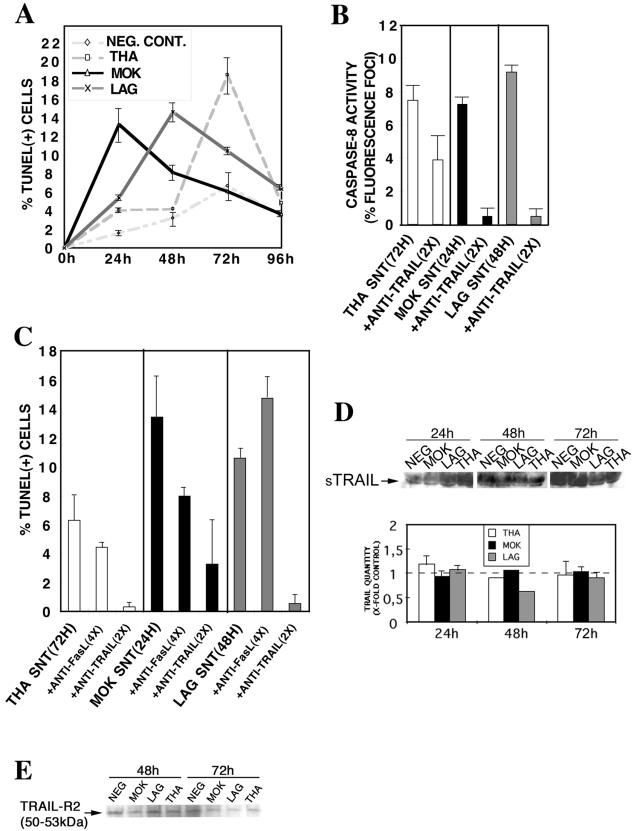
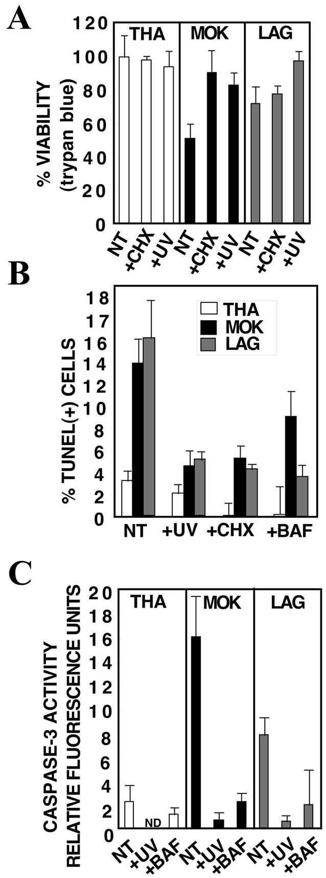
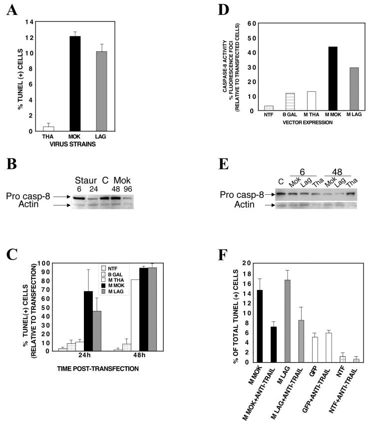
Lyssaviruses, which are members of the Rhabdoviridae family, induce apoptosis, which plays an important role in the neuropathogenesis of rabies. However, the mechanisms by which these viruses mediate neuronal apoptosis have not been elucidated. Here we demonstrate that the early induction of apoptosis in a model of lyssavirus-infected neuroblastoma cells involves a TRAIL-dependent pathway requiring the activation of caspase-8 but not of caspase-9 or caspase-10. The activation of caspase-8 results in the activation of caspase-3 and caspase-6, as shown by an increase in the cleavage of the specific caspase substrate in lyssavirus-infected cells. However, neither caspase-1 nor caspase-2 activity was detected during the early phase of infection. Lyssavirus-mediated cell death involves an interaction between TRAIL receptors and TRAIL, as demonstrated by experiments using neutralizing antibodies and soluble decoy TRAIL-R1/R2 receptors. We also demonstrated that the decapsidation and replication of lyssavirus are essential for inducing apoptosis, as supported by UV inactivation, cycloheximide treatment, and the use of bafilomycin A1 to inhibit endosomal acidification. Transfection of cells with the matrix protein induced apoptosis using pathways similar to those described in the context of viral infection. Furthermore, our data suggest that the matrix protein of lyssaviruses plays a major role in the early induction of TRAIL-mediated apoptosis by the release of a soluble, active form of TRAIL. In our model, Fas ligand (CD95L) appears to play a limited role in lyssavirus-mediated neuroblastoma cell death. Similarly, tumor necrosis factor alpha does not appear to play an important role.


Figures

FIG.1.
FIG.1.
Caspase activation in lyssavirus-infected neuroblastoma cells. Cells were infected with THA, MOK, or LAG at an MOI of 1. After incubation at 37°C for the indicated times, cells were processed. Results are expressed as means of data obtained in three independent experiments. Error bars, standard deviations. Significant effects are indicated by asterisks, pound signs, and plus signs (P < 0.05). (A) Percentages of lyssavirus-infected cells undergoing apoptosis and cell death according to the TUNEL assay and the amount of LDH released. (B) Caspase-1, -2, and -3 activity assays were evaluated by using Ac-DEVD-AMC-, VDVAD-pNA-, and Ac-YVAD-AMC-labeled substrates, respectively. (C) Caspase-6 activity was quantified at the single-cell level by using a medium containing a specific labeled peptide inhibitor (FAM-VEID-fmk) that recognizes the activated form of caspase-6. (D) Caspase-8 activity was quantified at the single-cell level by using a medium containing a specific labeled peptide inhibitor (FAM-LETD-fmk) that recognizes the activated form of caspase-8. LETDase activities were quantified in neuroblastoma cells infected at MOIs of 1 and 30. (E) Caspase-10 activity was quantified at the single-cell level by using a medium containing a specific labeled peptide inhibitor (FAM-AEVD-fmk) that recognizes the activated form of caspase-10. AEVDase activity was quantified in infected neuroblastoma cells (MOI, 30). (F) Western blot analysis of caspase-8 revealed the p54 fragment of the precursor form of caspase-8 (Pro casp-8). β-Actin was included as a loading control. NEG, negative control. (G) Caspase-9 activity was quantified at the single-cell level by using a medium containing a specific labeled peptide inhibitor (FAM-LEHD-fmk) that recognizes the activated form of caspase-9. LEHDase activities were quantified in neuroblastoma cells infected at MOIs of 1 and 30. (H) Cells were incubated in the presence or absence of a cell-permeant, irreversible pan-caspase inhibitor, Z-VAD-fmk, and a specific caspase-8 inhibitor, Z-IETD-fmk. CONTR, control.
FIG. 2.
FIG. 2.
Apoptosis and detection of caspase-8 activity in lyssavirus-infected neuroblastoma cells. Cells were either mock infected (A, E, and I) or infected with THA (B, F, and J), MOK (C, G, and K), or LAG (D, H, and L) (MOI, 1). (A through D) After 72 h at 37°C, cells were processed for TUNEL staining (red) and viral NC antigen staining (green) and were analyzed with a laser scanning confocal microscope (magnification, ×600). (E through H) Active caspase-8 was detected 48 h p.i. by using the carboxyfluorescein-labeled caspase inhibitor FAM-LETD-fmk (green). (I through L) Cells were counterstained with Hoechst 33342 (blue) and analyzed by fluorescence microscopy (magnification, ×1,000). (M) Matrix protein fused to His6 was visualized by use of an anti-His6 antibody conjugated with FITC (green). (N) Cells were transfected with the matrix-His6 fusion protein (M) and treated for the TUNEL assay (red). (O) Cells were counterstained with TOPRO-3 (blue) and analyzed by confocal microscopy (magnification, ×600).
FIG. 3.
FIG. 3.
TRAIL is necessary but not sufficient to activate caspase-8 and to induce apoptosis in lyssavirus-infected neuroblastoma cells. Neuroblastoma cells were pretreated with anti-TRAIL antibodies (2.75 to 5.5 μg/ml; 1× to 2×), soluble TRAIL receptors (22 μg/ml), anti-FasL antibodies (11 to 22 μg/ml; 4× to 8×), or anti-TNF-α antibodies (22 μg/ml; 8×) for 1 h before virus infection (MOI, 1). They were maintained in media containing anti-ligands for 72 h. Results are means of data obtained in two independent experiments. Error bars, standard deviations.Significant effects are indicated by asterisks and pound signs (P < 0.05). (A and B) The percentage of apoptotic cells was evaluated by the TUNEL assay (A) and caspase-8 activity (B). (C) FACS analysis of cytosolic and cell surface TRAIL expression using a purified rat anti-mouse TRAIL monoclonal antibody or a purified rat monoclonal immunoglobulin isotype control followed by an FITC-conjugated anti-rat antibody. Data were collected (10,000 events) by using a Becton Dickinson FACScalibur and were analyzed with CellQuest software. NT, not treated; CONT, control.
FIG. 4.
FIG. 4.
Induction in trans of apoptosis of lyssavirus-infected neuroblastoma cells is mediated by TRAIL through a synergistic released factor(s). Neuroblastoma cells were either mock infected (NEG. CONT.) or infected with THA, MOK, or LAG (MOI, 1). (A) At various times p.i., supernatants were filtered through a 100-kDa membrane and transferred to fresh, noninfected neuroblastoma cells. Graph shows the percentages of apoptotic nuclei in neuroblastoma cells following treatment with filtered supernatants taken from infected or mock-infected neuroblastoma cells. Times of supernatant harvest are given along the x axis. Apoptosis was assayed 72 h after supernatant transfer to noninfected cells. Results are means of data obtained in two independent experiments. Error bars, standard deviations. (B and C) Supernatants corresponding to maximum levels of apoptosis (with times of harvest given in parentheses) were incubated with an anti-mouse TRAIL neutralizing antibody (5.5 μg/ml; 2×). Caspase-8 activity (B) and TUNEL (C) assays were performed after 72 h of incubation with supernatants. (D) The release of the soluble form of TRAIL (sTRAIL) was analyzed by Western blotting of filter-concentrated supernatants at various times p.i. Protein concentrations of all samples were standardized by the BCA protein assay. Supernatants (40 μg) were subjected to SDS-polyacrylamide gel electrophoresis, blotted onto nitrocellulose membranes, and probed with anti-mouse TRAIL antibodies. (E) The amount of TRAIL-R2 receptor was analyzed by Western blotting using the solubilized cell membrane at 72 h p.i. Concentrated proteins (40 μg) were subjected to SDS-polyacrylamide gel electrophoresis, blotted, and probed with anti-human TRAIL-R2 antibodies.
FIG. 5.
FIG. 5.
Induction of cell death is dependent on viral decapsidation and replication. At 72 h p.i., the medium was removed, and cells were collected and counted under a microscope by using a hemacytometer chamber (A) or were treated for a TUNEL assay (B). Proteins were extracted and assayed for caspase-3 activity (C). Results are means of data obtained in three independent experiments. Error bars, standard deviations. (A) Virus inactivation by UV or pretreatment of neuroblastoma cells with CHX (10 μg/ml) protected neuroblastoma cells against MOK and to a lesser extent against LAG, as revealed by an increase in cell viability quantified by the trypan blue exclusion assay. (B and C) Pretreatment of neuroblastoma cells with BAF (10 μM), CHX (10 μg/ml), or UV-inactivated virus completely or partially protected them against LAG- or MOK-induced apoptosis, as evidenced by a decrease in the percentage of TUNEL-positive cells (B) and in caspase-3 activity (C). NT, not treated.
FIG. 6.
FIG. 6.
Matrix protein alone induces apoptosis by a TRAIL-dependent mechanism involving the caspase-8 pathway. (A) HeLa cells were infected with THA, MOK, or LAG (MOI, 15) and processed for the TUNEL assay. (B) Proteins (20 μg) were subjected to SDS-polyacrylamide gel electrophoresis, blotted, and probed with anti-human caspase-8. β-Actin was included as a loading control. (C through F) HeLa cells were either mock transfected (NTF) or transfected with pGFP (positive control), pM-GFP, or pM-(His)6. Cells were then processed for a TUNEL assay at the indicated time points (C), a caspase-8 activity assay at 24 h posttransfection (D), and Western blotting with anti-human caspase-8 (E). Cells were also processed for a neutralizing assay using an anti-human TRAIL neutralizing antibody at 48 h posttransfection (4.4 μg/ml) (F). Results are means of data obtained in two independent experiments. Error bars, standard deviations.

Similar articles

Cited by

References

    1. Adle-Biassette, H., H. Bourhy, M. Gisselbrecht, F. Chretien, L. Wingertsmann, M. Baudrimont, Y. Rotivel, B. Godeau, and F. Gray. 1996. Rabies encephalitis in a patient with AIDS: a clinicopathological study. Acta Neuropathol. 92:415-420. - PubMed
    1. Ashkenazi, A., and V. M. Dixit. 1999. Apoptosis control by death and decoy receptors. Curr. Opin. Cell Biol. 11:255-260. - PubMed
    1. Ashkenazi, A., and V. M. Dixit. 1998. Death receptors: signaling and modulation. Science 281:1305-1308. - PubMed
    1. Badrane, H., C. Bahloul, P. Perrin, and N. Tordo. 2001. Evidence of two Lyssavirus phylogroups with distinct pathogenicity and immunogenicity. J. Virol. 75:3268-3276. - PMC - PubMed
    1. Barker, V., G. Middleton, F. Davey, and A. M. Davies. 2001. TNF-α contributes to the death of NGF-dependent neurons during development. Nat. Neurosci. 4:1194-1198. - PubMed

Publication types