. 2004 Jun;78(12):6595-609.
doi: 10.1128/JVI.78.12.6595-6609.2004.

Adeno-associated virus type 2 VP2 capsid protein is nonessential and can tolerate large peptide insertions at its N terminus

Affiliations

Adeno-associated virus type 2 VP2 capsid protein is nonessential and can tolerate large peptide insertions at its N terminus

Kenneth H Warrington Jr et al. J Virol. 2004 Jun.

Abstract

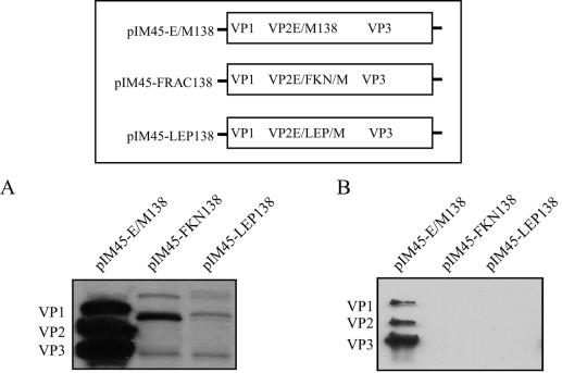
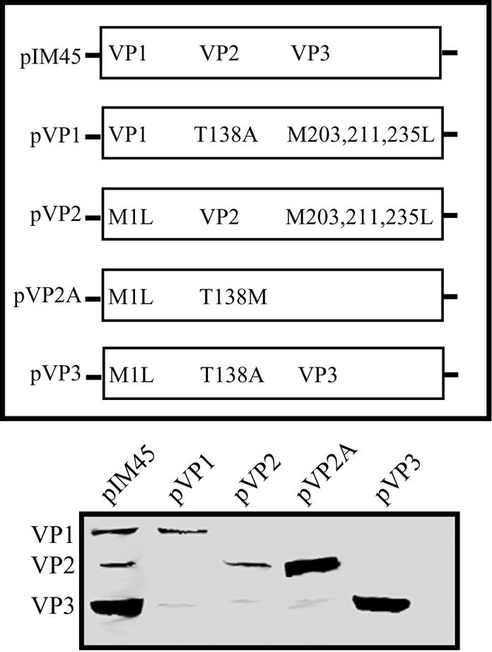
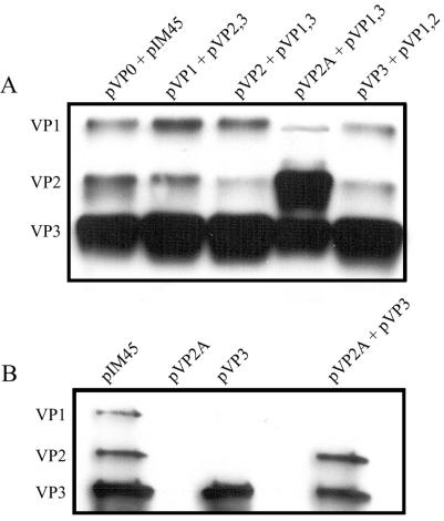
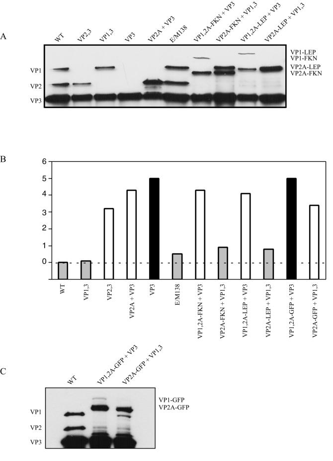
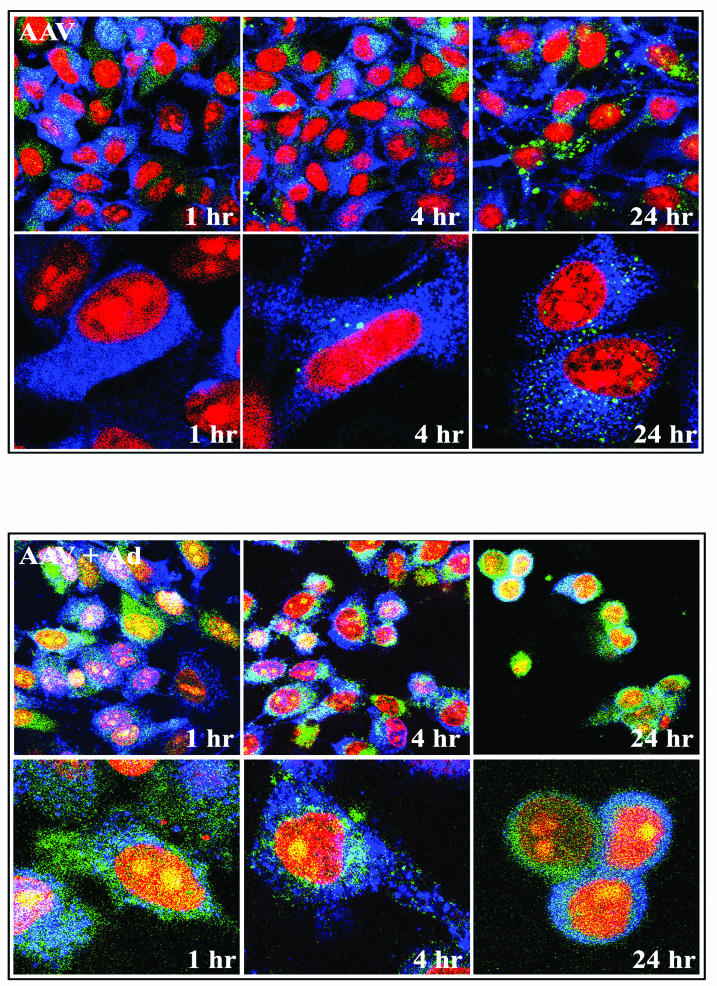
Direct insertion of amino acid sequences into the adeno-associated virus type 2 (AAV) capsid open reading frame (cap ORF) is one strategy currently being developed for retargeting this prototypical gene therapy vector. While this approach has successfully resulted in the formation of AAV particles that have expanded or retargeted viral tropism, the inserted sequences have been relatively short, linear receptor binding ligands. Since many receptor-ligand interactions involve nonlinear, conformation-dependent binding domains, we investigated the insertion of full-length peptides into the AAV cap ORF. To minimize disruption of critical VP3 structural domains, we confined the insertions to residue 138 within the VP1-VP2 overlap, which has been shown to be on the surface of the particle following insertion of smaller epitopes. The insertion of coding sequences for the 8-kDa chemokine binding domain of rat fractalkine (CX3CL1), the 18-kDa human hormone leptin, and the 30-kDa green fluorescent protein (GFP) after residue 138 failed to lead to formation of particles due to the loss of VP3 expression. To test the ability to complement these insertions with the missing capsid proteins in trans, we designed a system for producing AAV vectors in which expression of one capsid protein is isolated and combined with the remaining two capsid proteins expressed separately. Such an approach allows for genetic modification of a specific capsid protein across its entire coding sequence leaving the remaining capsid proteins unaffected. An examination of particle formation from the individual components of the system revealed that genome-containing particles formed as long as the VP3 capsid protein was present and demonstrated that the VP2 capsid protein is nonessential for viral infectivity. Viable particles composed of all three capsid proteins were obtained from the capsid complementation groups regardless of which capsid proteins were supplied separately in trans. Significant overexpression of VP2 resulted in the formation of particles with altered capsid protein stoichiometry. The key finding was that by using this system we successfully obtained nearly wild-type levels of recombinant AAV-like particles with large ligands inserted after residue 138 in VP1 and VP2 or in VP2 exclusively. While insertions at residue 138 in VP1 significantly decreased infectivity, insertions at residue 138 that were exclusively in VP2 had a minimal effect on viral assembly or infectivity. Finally, insertion of GFP into VP1 and VP2 resulted in a particle whose trafficking could be temporally monitored by using confocal microscopy. Thus, we have demonstrated a method that can be used to insert large (up to 30-kDa) peptide ligands into the AAV particle. This system allows greater flexibility than current approaches in genetically manipulating the composition of the AAV particle and, in particular, may allow vector retargeting to alternative receptors requiring interaction with full-length conformation-dependent peptide ligands.


Figures

FIG. 1.
FIG. 1.
Western blot analysis of AAV capsid proteins in 293 cell lysates (A) and iodixanol-purified virus stocks (B) following insertion of FKN or LEP peptides after residue 138 in the EagI-MluI cloning site engineered in the VP1-VP2 overlap region. Equal volumes of lysates or virus stocks were separated by SDS-10% polyacrylamide gel electrophoresis and analyzed by Western blotting with the B1 antibody. The diagram illustrates the position of the insertion of the E/M cloning site and the FKN and LEP ligands.
FIG. 2.
FIG. 2.
Mutants that express only two capsid proteins, showing Western blot analysis of capsids in cell lysates produced from 293 cells transfected with mutants that eliminate expression of one of the three AAV capsid proteins. Equal volumes of extracts were separated by SDS-10% polyacrylamide gel electrophoresis and analyzed by Western blotting with the B1 antibody. (A) The missense mutations within the start codons of the three capsid proteins (M1L, T138L, and M203L) are illustrated along with the capsid proteins expressed from each mutant on an SDS-acrylamide gel blotted with B1 antibody. (B) VP3-like proteins that result from readthrough translation. A mutation in the normal VP3 start codon produces a truncated capsid protein, VP3a; mutations in the first two methionines (pM203,211L) produce a second truncated protein, VP3b; and mutations in the first three methionines (pM203,211,235L; pVP1,2) eliminate all VP3-like proteins. (C) An alternative approach to eliminating VP3 expression while maximizing VP2 expression. pVP1,2A contains a standard ATG start codon for VP2 instead of ACG, a T138M mutation, thereby increasing VP2 expression and eliminating VP3 expression (compare pVP1,2A in panel C to pVP1,2 in panel B).
FIG. 3.
FIG. 3.
Mutants that express only a single capsid protein. Equal volumes of 293 cell extracts transfected with capsid mutants that express a single capsid protein were separated by SDS-10% polyacrylamide gel electrophoresis and analyzed by Western blotting with B1 antibody. The diagram illustrates the missense mutation(s) in each construct.
FIG. 4.
FIG. 4.
Which capsid mutants can make a virus particle? Western blot analysis of AAV virus purified by use of iodixanol step gradients as described in Materials and Methods following transfection of the indicated capsid mutants into 293 cells is shown. Equal volumes of the iodixanol fraction were separated by SDS-10% polyacrylamide gel electrophoresis and analyzed by Western blotting with B1 antibody. (A) Effect of the M203L, M211L, and M235L mutations on particle formation. (B) Particle formation from mutants that lack a specific capsid protein. (C) Particle formation from mutants that express a single capsid protein.
FIG. 5.
FIG. 5.
Complementation of mutants that make a single capsid protein. (A) Western blot analysis of AAV particles purified by use of iodixanol step gradients and heparin column chromatography following transfection of 293 cells with the complementation groups described in Table 3. (B) Western blot analysis of iodixanol fractions of particles obtained from transfection with pVP2A, pVP3, or both plasmids. Equal volumes of purified virus stocks were separated by SDS-10% acrylamide gel electrophoresis and analyzed by Western blotting with the B1 antibody.
FIG. 6.
FIG. 6.
Capsid complementation strategy for creating particles with large peptide insertions in the VP1-VP2 overlap region, showing Western blotting of equal volumes of iodixanol stocks of AAV-like particles containing FKN or LEP insertions at position 138. (A) Diagram of constructs used to complement insertions at amino acid 138 in both VP1 and VP2A or just VP2A. (B) Particles with the FKN insertion were purified by use of iodixanol gradients and probed on SDS-10% polyacrylamide gels with anticapsid (B1) antibody or anti-FKN antibody. (C) Particles with the LEP insertion were purified by use of iodixanol gradients and probed on SDS-10% polyacrylamide gels with anticapsid (B1) antibody or anti-LEP antibody.
FIG. 7.
FIG. 7.
Capsid protein stoichiometry and infectivity of AAV virus stocks missing a capsid protein or containing a ligand insertion. (A) Western blot of virus stocks purified by use of iodixanol gradients and heparin sulfate column chromatography. Approximately 1011 AAV-like particles were separated by SDS-10% polyacrylamide gel electrophoresis and analyzed by Western blotting with the B1 antibody. WT, wild type. (B) Particle-to-infectivity ratios of AAV-like particles relative to that of pIM45. The particle-to-infectivity ratio for each particle was calculated by dividing the average genomic titer by the average FCA titer (see Material and Methods and Table 2). The particle-to-infectivity ratio for each type of virus was then normalized to that of wild-type virus (pIM45) by dividing the particle-to-infectivity of each AAV-like particle by the particle-to-infectivity of pIM45, and the log10 value of the ratio was plotted. The wild-type pIM45 ratio equals zero and is indicated by the dashed line. Grey bars, particles with infectivity comparable to that of pIM45 (within 1 log unit); white bars, particles with significantly reduced infectivity (1- to 4-log-unit-lower infectivity); black bars, particles that were essentially noninfectious (>4-log-unit-lower infectivity). (C) Western blot of approximately 1011 AAV-like particles with GFP inserted in the capsid. Virus samples were purified as for panel A above, fractionated by SDS-10% polyacrylamide gel electrophoresis, and analyzed by Western blotting with the B1 antibody.
FIG. 8.
FIG. 8.
Time course of VP1,2A-GFP plus VP3 particle trafficking following infection in the absence (top panels) and presence (bottom panels) of Ad5. HeLa cells were infected with AAV containing a GFP insertion at an MOI of 10,000 with or without Ad5 at an MOI of 20. Vectors remained on the cells for the duration of the time course. The input capsids appear green from the native GFP fluorescence of the capsid, the nuclei are stained red with PI, and early endosomal antigen 1 is stained blue.

References

    1. Becerra, S. P., F. Koczot, P. Fabisch, and J. A. Rose. 1988. Synthesis of adeno-associated virus structural proteins requires both alternative mRNA splicing and alternative initiations from a single transcript. J. Virol. 62:2745-2754. - PMC - PubMed
    1. Becerra, S. P., J. A. Rose, M. Hardy, B. M. Baroudy, and C. W. Anderson. 1985. Direct mapping of adeno-associated virus capsid proteins B and C: a possible ACG initiation codon. Proc. Natl. Acad. Sci. USA 82:7919-7923. - PMC - PubMed
    1. Bowles, D. E., J. E. Rabinowitz, and R. J. Samulski. 2003. Marker rescue of adeno-associated virus (AAV) capsid mutants: a novel approach for chimeric AAV production. J. Virol. 77:423-432. - PMC - PubMed
    1. Buller, R. M., and J. A. Rose. 1978. Characterization of adenovirus-associated virus-induced polypeptides in KB cells. J. Virol. 25:331-338. - PMC - PubMed
    1. Buning, H., M. U. Ried, L. Perabo, F. M. Gerner, N. A. Huttner, J. Enssle, and M. Hallek. 2003. Receptor targeting of adeno-associated virus vectors. Gene Ther. 10:1142-1151. - PubMed

Publication types