

Role of an arbovirus nonstructural protein in cellular pathogenesis and virus release
- PMID: 15163755
- PMCID: PMC416502
- DOI: 10.1128/JVI.78.12.6649-6656.2004
Role of an arbovirus nonstructural protein in cellular pathogenesis and virus release
Abstract
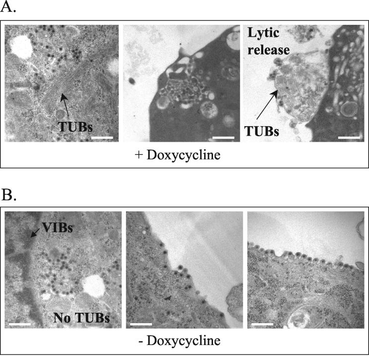
The insect-borne Bluetongue virus (BTV) is considered the prototypic Orbivirus, a member of the Reovirus family. One of the hallmarks of Orbivirus infection is the production of large numbers of intracellular tubular structures of unknown function. For BTV these structures are formed as the polymerization product of a single 64-kDa nonstructural protein, NS1, encoded by the viral double-stranded RNA genome segment 6. Although the NS1 protein is the most abundant viral protein synthesized in infected cells, its function has yet to be determined. One possibility is that NS1 tubules may be involved in the translocation of newly formed viral particles to the plasma membrane, and NS1-specific monoclonal antibodies have been shown to react with viral particles leaving infected cells. In the present study we generated a mammalian cell line that expresses a recombinant single-chain antibody fragment (scFv) derived from an NS1-specific monoclonal antibody (10B1) and analyzed the effect that this intracellular antibody has on BTV replication. Normally, BTV infection of mammalian cells in culture results in a severe cytopathic effect within 24 to 48 h postinfection manifested by cell rounding, apoptosis, and lytic release of virions into the culture medium. However, infection of scFv-expressing cells results in a marked reduction in the stability of NS1 and formation of NS1 tubules, a decrease in cytopathic effect, an increased release of infectious virus into the culture medium, and budding of virions from the plasma membrane. These results suggest that NS1 tubules play a direct role in the cellular pathogenesis and morphogenesis of BTV.
Figures
Similar articles
-
A viral nonstructural protein regulates bluetongue virus trafficking and release.J Virol. 2009 Jul;83(13):6806-16. doi: 10.1128/JVI.00263-09. Epub 2009 Apr 15. J Virol. 2009. PMID: 19369335 Free PMC article.
-
Bluetongue virus non-structural protein 1 is a positive regulator of viral protein synthesis.Virol J. 2012 Aug 29;9:178. doi: 10.1186/1743-422X-9-178. Virol J. 2012. PMID: 22931514 Free PMC article.
-
Analysis of murine B-cell epitopes on bluetongue virus 12 nonstructural protein 1.Appl Microbiol Biotechnol. 2015 Feb;99(3):1309-21. doi: 10.1007/s00253-014-6150-4. Epub 2014 Oct 26. Appl Microbiol Biotechnol. 2015. PMID: 25343975
-
Genetically engineered multi-component virus-like particles as veterinary vaccines.Immunol Cell Biol. 1993 Oct;71 ( Pt 5):381-9. doi: 10.1038/icb.1993.44. Immunol Cell Biol. 1993. PMID: 8270267 Review.
-
Bluetongue virus assembly and exit pathways.Adv Virus Res. 2020;108:249-273. doi: 10.1016/bs.aivir.2020.08.002. Epub 2020 Sep 16. Adv Virus Res. 2020. PMID: 33837718 Free PMC article. Review.
Cited by
-
Collective fusion activity determines neurotropism of an en bloc transmitted enveloped virus.Sci Adv. 2023 Jan 27;9(4):eadf3731. doi: 10.1126/sciadv.adf3731. Epub 2023 Jan 27. Sci Adv. 2023. PMID: 36706187 Free PMC article.
-
Clinical, Virological, and Immunological Features in Cosmopolitan Genotype DENV-2-Infected Patients during a Large Dengue Outbreak in Sri Lanka in 2017.Am J Trop Med Hyg. 2023 Sep 11;109(4):917-925. doi: 10.4269/ajtmh.22-0780. Print 2023 Oct 4. Am J Trop Med Hyg. 2023. PMID: 37696512 Free PMC article.
-
Studies on the clinical symptoms, virus distribution, and mRNA expression of several antiviral immunity-related genes in grass carp after infection with genotype II grass carp reovirus.Arch Virol. 2020 Jul;165(7):1599-1609. doi: 10.1007/s00705-020-04654-y. Epub 2020 May 12. Arch Virol. 2020. PMID: 32399788
-
Identification of Orbivirus Non-Structural Protein 5 (NS5), Its Role and Interaction with RNA/DNA in Infected Cells.Int J Mol Sci. 2023 Apr 6;24(7):6845. doi: 10.3390/ijms24076845. Int J Mol Sci. 2023. PMID: 37047816 Free PMC article.
-
Comparative Virus-Host Protein Interactions of the Bluetongue Virus NS4 Virulence Factor.Viruses. 2022 Jan 19;14(2):182. doi: 10.3390/v14020182. Viruses. 2022. PMID: 35215776 Free PMC article.
References
-
- Blaney, J. E., Jr., G. G. Manipon, B. R. Murphy, and S. S. Whitehead. 2003. Temperature sensitive mutations in the genes encoding the NS1, NS2A, NS3, and NS5 nonstructural proteins of dengue virus type 4 restrict replication in the brains of mice. Arch Virol. 148:999-1006. - PubMed
-
- Cormack, B. P., R. H. Valdivia, and S. Falkow. 1996. FACS-optimized mutants of the green fluorescent protein (GFP). Gene 173:33-38. - PubMed
-
- Eaton, B. T., A. D. Hyatt, and S. M. Brookes. 1990. The replication of bluetongue virus. Curr. Top. Microbiol. Immunol. 162:89-118. - PubMed
Publication types
MeSH terms
Substances
Grants and funding
LinkOut - more resources
Full Text Sources
