. 2004 Jun;52(5):1255-69.
doi: 10.1111/j.1365-2958.2004.04063.x.

Screening for synthetic lethal mutants in Escherichia coli and identification of EnvC (YibP) as a periplasmic septal ring factor with murein hydrolase activity

Affiliations

Screening for synthetic lethal mutants in Escherichia coli and identification of EnvC (YibP) as a periplasmic septal ring factor with murein hydrolase activity

Thomas G Bernhardt et al. Mol Microbiol. 2004 Jun.

Abstract

Bacterial cytokinesis is driven by the septal ring apparatus, the assembly of which in Escherichia coli is directed to mid-cell by the Min system. Despite suffering aberrant divisions at the poles, cells lacking the minCDE operon (Min(-)) have an almost normal growth rate. We developed a generally applicable screening method for synthetic lethality in E. coli, and used it to select for transposon mutations (slm) that are synthetically lethal (or sick) in combination with DeltaminCDE. One of the slm insertions mapped to envC (yibP), proposed to encode a lysostaphin-like, metallo-endopeptidase that is exported to the periplasm by the general secretory (Sec) pathway. Min(-) EnvC(-) cells showed a severe division defect, supporting a role for EnvC in septal ring function. Accordingly, we show that an EnvC-green fluorescent protein fusion, when directed to the periplasm via the twin-arginine export system, is both functional and part of the septal ring apparatus. Using an in-gel assay, we also present evidence that EnvC possesses murein hydrolytic activity. Our results suggest that EnvC plays a direct role in septal murein cleavage to allow outer membrane constriction and daughter cell separation. By uncovering genetic interactions, the synthetic lethal screen described here provides an attractive new tool for studying gene function in E. coli.


Figures

Fig. 1
Fig. 1
Colony sectoring and the screen for slm mutants. Colony phenotypes of strain TB15/pTB8 [ΔlacIZYA ΔminCDE/Plac::minCDE lacZ] (left) and a slm11 derivative (right) grown on non-selective LB agar supplemented with 500 μM IPTG and 60 μg ml−1 Xgal. Note that slm+ cells produced both solid-white and sectored-blue colonies with an asterisk-like appearance, indicating rapid loss of pTB8, whereas slm11 colonies were almost exclusively solid-blue, indicating selective pressure to retain the plasmid. A. Typical sectored (slm+) and solid-blue (slm11) colonies from (B) in more detail. Although not visible here, the slm11 strain also produced some tiny white colonies, consisting of highly filamentous cells.
Fig. 2
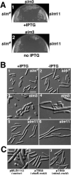
Fig. 2. Growth and morphology of the slm mutants
A. Growth phenotypes of TB15/pTB8 [ΔlacIZYA ΔminCDE/Plac::minCDE lacZ] (left) and its slm3 (centre) and slm11 (right) derivatives on LB agar with (1) or without (2) 500 μM IPTG. Note the poor growth of the slm3 and slm11 mutants in the absence of IPTG. B. DIC micrographs of TB15/pTB8 (1 and 2) and its slm3 (3 and 4) and slm11 (5 and 6) derivatives grown in liquid LB with (1, 3 and 5) or without (2, 4 and 6) 500 μM IPTG. Cells were grown to an OD600 of 0.6–0.7 and fixed. Bar equals 5 μm. C. Correction of the slm3 phenotype by multicopy pbpA rodA. DIC micrographs of strain TB81 [slm3 Para::minCDE] carrying pMLB1113 [vector] (1), pTB58 [Plac::ybeBA pbpA rodA] (2) or pTB59 [Plac::pbpA rodA] (3). Cells were grown in LB with no (1 and 2) or 50 μM (3) IPTG to an OD600 of 0.6–0.8 and fixed. Note that cells were Min because arabinose was omitted from the medium. Bar equals 2 μm.
Fig. 3
Fig. 3
Maps of plasmids, phage constructs and positions of the transposon insertions. Diagrams of the ybeBA pbpA rodA (A) and envC (B) loci are shown, indicating the location and orientation of the EZTnKan-2 insertions responsible for the Slm phenotype. The SWISSPROT annotations for YbeB (P05848) and EnvC (P37690) were used to construct the figure. Plasmid and phage inserts (solid lines), the lac or phage T7 promoters (arrows), gfpmut2 (grey box), the FKH tag (white box), the insertion of the AmiC signal sequence coding region (ssamiC) and the presence of a T7 gene10 ribosome binding site (*) are indicated. The predicted domain organization of EnvC is shown in (B). The signal sequence (SS), predicted coiled-coil region (CC) and M37 metallo-endopeptidase domain (M37) are shown. The relevant amino acid residue numbers for each domain are given above the diagram. The numbers were derived from Hara et al. (2002) but modified to be consistent with the SWISSPROT annotation.
Fig. 4
Fig. 4
Septal ring and nucleoid positioning in EnvC and EnvC Min mutants. Micrographs show representative fields of TB28(λCH151) [wt (Plac::zipA-gfp)] (A), TB35(λCH151) [ΔenvC::aph (Plac::zipA-gfp)] (B), TB57(λCH151) [Para::minCDE (Plac::zipA-gfp)] (C) and TB58(λCH151) [ΔenvC::aph Para::minCDE (Plac::zipA-gfp)] (D) cells grown in LB with 50 μM IPTG to an OD600 of 0.6. The cells were fixed, stained with DAPI and viewed for GFP fluorescence (1, pseudocoloured green), DAPI fluorescence (2, pseudocoloured red) and by DIC (4). (3) Overlays of the GFP and DAPI channels. Single arrowheads point to ZipA–GFP rings forming in regions with significant DAPI staining, and double arrowheads point to aberrant ZipA–GFP structures. The arrow in (B) points to a cell constriction without an associated ZipA–GFP ring. Bar equals 2 μm.
Fig. 5
Fig. 5. Measurement of cell length and ZipA–GFP ring formation in EnvC cells
A. Cell lengths and ZipA–GFP ring distributions were measured for 197 TB28(λCH151) [wt (Plac::zipA-gfp)] cells and 181 TB35(λCH151) [ΔenvC::aph (Plac::zipA-gfp)] cells from the experiment described in Fig. 4. The average cell length and length per ZipA–GFP ring (L/R ratio) are given. The types of cells observed were separated into seven classes: (1) unconstricted cells without a ring; (2) unconstricted cells with a medial ring; (3) medially constricting cells with a medial ring; (4) medially constricting cells with no ring; (5) medially constricting cells with one ring at a quarter position; (6) medially constricting cells with a ring at each quarter position; and (7) other types of cells. The ZipA–GFP rings in cells of class 5 tended to be very dim, suggesting that they just began to form. Many cells in class 6 had very shallow constrictions at the quarter positions. The percentage of each cell class observed is indicated to the right of the cartoons. Note that the cartoons do not reflect the relative sizes of the cells in each class. EnvC cells in classes 3 and 6 were often twice as long as the typical wild-type cells in class 3. B. Histograms indicating the number of cells with a particular L/R ratio. This ratio was used to normalize for the high frequency of TB35(λCH151) with two rings. The data from (A), excluding the minority of cells without rings, were used to generate the histograms.
Fig. 6
Fig. 6. EnvC is recruited to the septal ring
A. Immunoblot probed with anti-GFP antibodies. Lanes contained whole-cell extracts of TB28(λTB47) [wt (Plac::ssamiC-envC-gfp)] (lane 1) or TB44(λTB47) [ΔenvC::frt (Plac::ssamiC-envC-gfp)] (lane 2). Extracts were prepared from cells grown to an OD600 of 0.5–0.6 in minimal medium supplemented with 50 μM IPTG. The arrow indicates the position of TTEnvC–GFP. Numbers on the left (in kDa) indicate the positions of molecular weight markers. B. Release of TTEnvC–GFP from spheroplasts. TB28(λTB47) [wt (Plac::ssamiC-envC-gfp)] was grown in LB supplemented with 0.2% maltose and 50 μM IPTG. One aliquot of cells was used to prepare a total-cell extract. The remaining cells were converted to spheroplasts and pelleted by centrifugation. The resulting pellet (P) and supernatant (S) fractions, along with the total-cell extract (T), were analysed by SDS–PAGE and immunoblotting for GFP, FtsZ or MalE as indicated. FtsZ and MalE served as markers for the cytoplasm and periplasm respectively. C. GFP fluorescence (1–6) and DIC (1′-6′) micrographs of live TB28(λTB47) [wt (Plac::ssamiC-envC-gfp)] (1 and 2), TB44(λTB47) [ΔenvC::frt (Plac::ssamiC-envC-gfp)] (3), MC4100(λTB47) [wt (Plac::ssmiC-envC-gfp)] (4), B1LKO(λTB47) [ΔtatC (Plac::ssamiC-envC-gfp)] (5) and TB28(λTB47)/pJE80 [wt (Plac::ssamiC-envC-gfp)/Para::sfiA] (6) cells grown as in (A), except that the culture used for (6) contained 0.2% arabinose and 100 μM IPTG. In (1), the single arrow points to a deeply constricted cell with a bright focus of EnvC–GFP at the septum, and the double arrow points to a cell with a shallow constriction that has a bright EnvC–GFP ring as well as a peripheral signal. In (2), the single arrow points to a small cell with an apparent accumulation of EnvC–GFP signal at mid-cell, and the double arrow points to a very deeply constricted cell with a dominant peripheral signal. This cell has presumably just completed division, and the daughters are about to separate. Bar equals 2 μm.
Fig. 7
Fig. 7
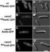
Evidence for an increased periplasmic volume at the septa of EnvC cells. GFP fluorescence (A–D) and DIC (A′–D′) micrographs show representative live cells of TB28(λTB46) [wt (Plac::ssamiC-gfp)] (A), TB44(λTB46) [ΔenvC::frt (Plac::ssamiC-gfp)] (B), TB44/pTB32 [ΔenvC::frt/Plac::amiA-gfp)] (C) and TB44(λTB6) [ΔenvC::frt (Plac::sstorA-gfp)] (D) grown in minimal medium supplemented with 500 (A and B), 250 (C) or 50 μM (D) IPTG. Cells were grown to an OD600 of 0.5–0.6. Bar equals 2 μm.
Fig. 8
Fig. 8
EnvC has murein hydrolytic activity. Coomassie-stained gel (A) and methylene blue-stained zymogram (B) contained molecular weight markers (in descending order: 66, 45, 36, 29, 24, 20 and 14 kDa) (lane 1), 5 μg of BSA (lane 2), 5 μg of egg white lysozyme (Lys) (lane 3) and 5.0, 2.5 or 1.3 μg of purified EnvC–FKH (lanes 4–6). Lys and EnvC–FKH yielded significant clear zones in the zymogram. Minor species running below full-length EnvC–FKH also promoted some clearing. These were probably EnvC–FKH fragments that retained some activity.

References

    1. Abanes-De Mello A, Sun YL, Aung S, Pogliano K. A cytoskeleton-like role for the bacterial cell wall during engulfment of the Bacillus subtilis forespore. Genes Dev. 2002;16:3253–3264. - PMC - PubMed
    1. Åkerlund T, Bernander R, Nordström K. Cell division in Escherichia coli minB mutants. Mol Microbiol. 1992;6:2073–2083. - PubMed
    1. Bateman A, Birney E, Cerruti L, Durbin R, Etwiller L, Eddy SR, et al. The Pfam protein families data-base. Nucleic Acids Res. 2002;30:276–280. - PMC - PubMed
    1. Begg K, Nikolaichik Y, Crossland N, Donachie WD. Roles of FtsA and FtsZ in activation of division sites. J Bacteriol. 1998;180:881–884. - PMC - PubMed
    1. Bender A, Pringle JR. Use of a screen for synthetic lethal and multicopy suppressee mutants to identify two new genes involved in morphogenesis in Saccharomyces cerevisiae. Mol Cell Biol. 1991;11:1295–1305. - PMC - PubMed

Publication types

MeSH terms

LinkOut - more resources