Review
. 2004 Jun;12(6):279-87.
doi: 10.1016/j.tim.2004.04.005.

Norovirus disease: changing epidemiology and host susceptibility factors

Affiliations
Review

Norovirus disease: changing epidemiology and host susceptibility factors

Anne M Hutson et al. Trends Microbiol. 2004 Jun.

Abstract

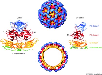
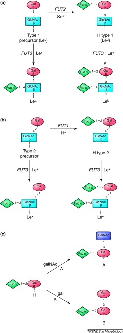
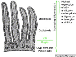
Noroviruses cause the majority of acute viral gastroenteritis cases that occur worldwide. The increased recognition of noroviruses as the cause of outbreaks and sporadic disease is due to the recent availability of improved norovirus-specific diagnostics. Transmission of these viruses is facilitated by their high prevalence in the community, shedding of infectious virus particles from asymptomatic individuals and the high stability of the virus in the environment. Currently, the spectrum of clinical disease and the understanding of host susceptibility factors are changing. Cases of chronic norovirus gastroenteritis have been observed in transplant recipients and unusual clinical presentations have been recognized in otherwise healthy adults that are under physical stress. Recently, noroviruses were found to bind to gut-expressed carbohydrates, leading to a correlation between a person's genetically determined carbohydrate expression and their susceptibility to Norwalk virus infection. Greater community surveillance and further investigation of carbohydrate receptor-binding properties could provide further insights into norovirus transmission, susceptibility and pathogenesis, and should aid in developing vaccines and antiviral therapies for this common viral disease.


Figures

Figure 1
Figure 1
The Norwalk virus-like particle (NV VLP) structure has been solved by cryo-electron microscopic reconstruction to 22 Å (top, surface representation; bottom, cross-section) and by x-ray crystallography to 3.4 Å. The NV VLPs have 90 dimers of capsid protein (left, ribbon diagram) assembled in T=3 icosahedral symmetry. Each monomeric capsid protein (right, ribbon diagram) is divided into an N-terminal arm region (green) facing the interior of the VLP, a shell domain (S-domain, yellow) that forms the continuous surface of the VLP, and a protruding domain (P-domain) that emanates from the S-domain surface. The P-domain is further divided into subdomains, P1 and P2 (red and blue, respectively) with the P2-subdomain at the most distal surface of the VLPs. Adapted, with permission, from Refs. , .
Figure 2
Figure 2
ABH and Lewis antigens are synthesized by sequential enzymatic transfer of single carbohydrate residues to specific precursor carbohydrate substrates. (a,b) H antigens are made by enzymatic addition of a fucose (Fuc) residue to the terminal galactose (Gal) residue in α1,2 linkage. (a) Secretor positive (Se+) individuals express the FUT2 gene product, a fucosyltransferase that adds Fuc to a type 1 precursor to make H type 1 (also known as Lewis(d) or Led). Eighty percent of Northern Europeans and Caucasian Americans are Se+. (b) The FUT1 fucosyltransferase adds Fuc to a type 2 precursor to make H type 2. Less than 0.002% of people throughout the world lack FUT1 expression; they also do not express H antigen on their red blood cells (histo-blood group type Bombay), which is normally expressed on type O red blood cells. Type 1 and 2 precursor substrates have different Gal to N-acetylglucosamine (GlcNAc) linkages, (a) Galβ1,3-GlcNAcβ- and (b) Galβ1,4-GlcNAcβ-, respectively. The Lewis carbohydrate antigens are made when the FUT3 enzyme is expressed in Lewis positive (Le+) individuals. Eighty percent of Northern Europeans and Caucasian Americans individuals are Le+, independent of secretor status. (a,b)FUT3 transfers Fuc to the GlcNAc of type 1 and 2 precursors and H types 1 and 2 in α1,4 and α1,3 linkages, respectively. (c) H types 1 and 2 are the terminal moieties expressed in histo-blood group type O individuals, but in types A, B and AB individuals the H antigens are further modified by enzymes that transfer N-acetylgalactosamine (GalNAc, type A), Gal (type B), or either carbohydrate (type AB) to the terminal Gal residue of an H antigen in α1,3 linkage. ABH, Lewis and secretor phenotypes and enzymatic pathways are described in greater detail in other reviews , .
Figure 3
Figure 3
The small intestinal gut section shows the villi projecting into the lumen (top) with the crypts beneath the villi. Within the crypts are the progenitor stem cells of the small intestine. Crypt stem cell division supplies cells that differentiate as they are pushed down into the base of the crypt, becoming paneth cells, or up into the villus, becoming goblet cells or enterocytes. Paneth cells at the base of the crypt secrete lysozyme, goblet cells secrete mucins, undifferentiated enterocytes secrete chloride ions, and differentiated enterocytes absorb nutrients from the gut lumen until they undergo apoptosis and are sloughed off the villus. As enterocytes differentiate and travel up the villus, they express higher amounts and a greater variety of carbohydrates, including ABH and Lewis histo-blood group antigens . Further description of these and other specialized intestinal cells can be found elsewhere .

References

    1. Dolin R. Biological properties of Norwalk agent of acute infectious nonbacterial gastroenteritis. Proc. Soc. Exp. Biol. Med. 1972;140:578–583. - PubMed
    1. Adler J.L, Zickl R. Winter vomiting disease. J. Infect. Dis. 1969;119:668–673. - PubMed
    1. Kapikian A.Z. Visualization by immune electron microscopy of a 27 nm particle associated with acute infectious nonbacterial gastroenteritis. J. Virol. 1972;10:1075–1081. - PMC - PubMed
    1. Green K.Y. Human calciviruses. In: Knipe D.M, editor. Vol. 1. Lippincott, Williams & Wilkins; 2001. pp. 841–874. (Fields Virology).
    1. Jiang X. Norwalk virus genome cloning and characterization. Science. 1990;250:1580–1583. - PubMed

Publication types

MeSH terms

Substances