

doi: 10.1126/science.1096706.
A family with severe insulin resistance and diabetes due to a mutation in AKT2
Affiliations
- PMID: 15166380
- PMCID: PMC2258004
- DOI: 10.1126/science.1096706
Item in Clipboard
A family with severe insulin resistance and diabetes due to a mutation in AKT2
Science.
.
Abstract
Inherited defects in signaling pathways downstream of the insulin receptor have long been suggested to contribute to human type 2 diabetes mellitus. Here we describe a mutation in the gene encoding the protein kinase AKT2/PKBbeta in a family that shows autosomal dominant inheritance of severe insulin resistance and diabetes mellitus. Expression of the mutant kinase in cultured cells disrupted insulin signaling to metabolic end points and inhibited the function of coexpressed, wild-type AKT. These findings demonstrate the central importance of AKT signaling to insulin sensitivity in humans.
Figures
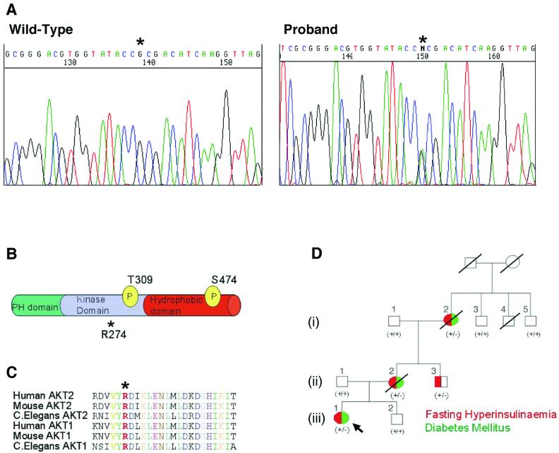
Detection of a non-conservative heterozygous mutation in AKT2 that co-segregates with severe insulin resistance. (A) Direct sequencing of genomic DNA from the proband, subject (iii)/1 (right) and a control subject (left). Asterisk indicates the heterozygous G to A substitution that produces H274. (B) Location of R274 in relation to known functional domains and phosphorylation sites required for activation of AKT2. (C) R274 (in red and marked *) is highly conserved across different AKT isoforms and diverse species. (D) Family pedigree demonstrating co-segregation of clinical phenotype (also see Table S1) with the R274H mutation. All family members heterozygous for the mutation (+/-) are hyperinsulinemic and 3 of 4 have diabetes mellitus. All wild type subjects (+/+) are normoinsulinemic and non-diabetic. Red denotes fasting hyperinsulinemia. Green denotes Diabetes Mellitus.
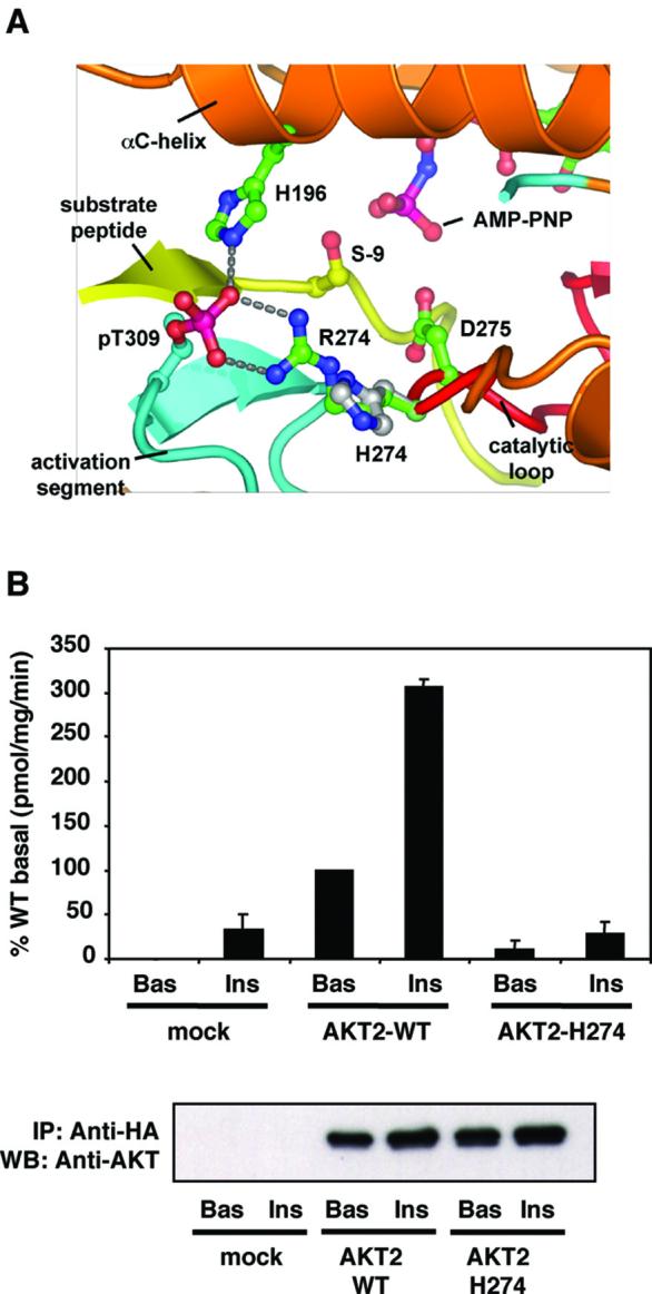
The substitution of R274 by histidine disrupts the kinase domain and abolishes AKT2 kinase activity. (A) Proposed effects of R274H on a structural model of the AKT2 kinase domain. In the wild type protein, R274 contacts phosphoT309 (pT309), organising the activation segment to place the substrate peptide correctly for catalysis. Substitution of H for R274 is predicted to disrupt the conformation of both the activation segment and catalytic loops. (B) HA-AKT2 and HA-AKT2H274 were immunoprecipitated from lysates of appropriately transfected CHO-T cells and enzyme activity was determined by an in vitro kinase assay (upper panel). Mock-transfected cells received empty vector only. Cells were treated without (Bas) or with (Ins) 100nM insulin for 10 min prior to lysis. Data are means±SEM of 4 experiments, Crosstide was used as the substrate. Duplicate immunoprecipitates were also immunoblotted with anti-AKT2 to demonstrate equal expression of the wild type and mutant proteins (lower panel).
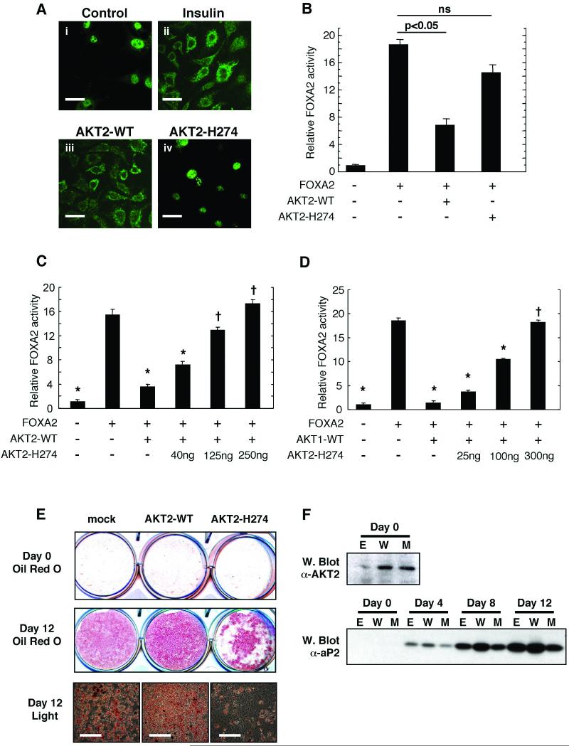
Functional properties of wild type AKT2 and AKT2H274 in cultured human liver and rodent fat cells (A) HepG2 cells were treated in the absence (panel i) or presence (panel ii) of 50nM insulin, fixed and probed with anti-FOXA2 antibodies to determine intracellular localization (7). The same assay was performed with cells transfected with wild type AKT2 (panel iii) or AKT2H274 (panel iv). Scale bars (40μm each) are shown in white. (B) Luciferase activity was determined in extracts of HepG2 cells transfected with pPCK1 reporter construct with or without FOXA2 in the absence or presence of wild type AKT2 or AKT2H274 (ns denotes no significant difference). Alternatively HepG2 cells were transfected with pPCK1 reporter construct with or without FOXA2 and either wild type AKT2 (125ng) (C) or wild type AKT1 (100ng) (D). In each case AKT2H274 was co-transfected in increasing quantities as indicated. * indicates significant difference from activity in cells transfected with FOXA2 alone (p<0.05). † denotes no significant difference in this comparison. In all cases data are means±SD of four experiments and all data was normalized to co-expressed β-galactosidase activity. (E) 3T3-L1 preadipocytes were stably transfected with empty vector (mock), wild type AKT2 or AKT2H274. 2-day post confluent cells (Day 0) or cells induced to differentiate for 12 days were fixed and stained with oil red O to assess lipid accumulation. Images of day 12 differentiated cells were also obtained by light microscopy (lower panels) and scale bars (200μm each) are shown in white. (F) Lysates were prepared from cells transfected with empty vector (E) wild type AKT2 (W) of AKT2H274 (M) at 2 days post-confluence (Day 0) or at various intervals up to day 12 post-induction of differentiation. Day 0 samples were immunoblotted for AKT2 expression and all lysates were immunoblotted for aP2 expression.
References
Publication types
MeSH terms
Substances
Associated data
- Actions
Grants and funding
LinkOut - more resources
Full Text Sources
Other Literature Sources
Medical
Molecular Biology Databases
Miscellaneous
