Comparative Study
. 2004 Apr;69(4 Pt 1):040901.
doi: 10.1103/PhysRevE.69.040901. Epub 2004 Apr 30.

Diffuse scattering provides material parameters and electron density profiles of biomembranes

Affiliations
Comparative Study

Diffuse scattering provides material parameters and electron density profiles of biomembranes

Yufeng Liu et al. Phys Rev E Stat Nonlin Soft Matter Phys. 2004 Apr.

Abstract

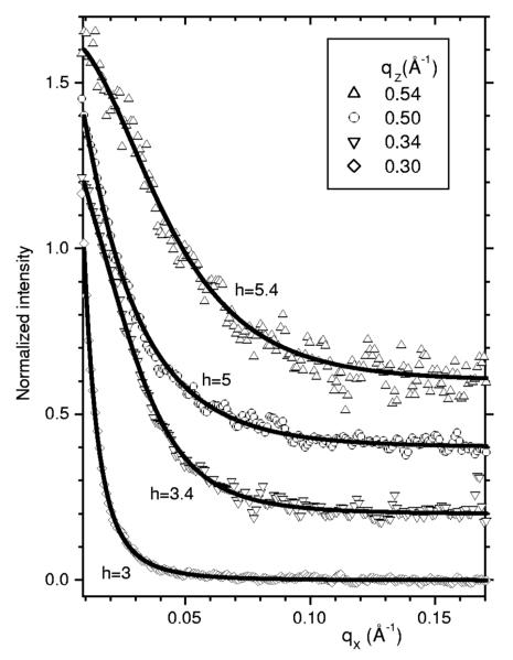
Fully hydrated stacks of DOPC lipid bilayer membranes generate large diffuse x-ray scattering that corrupts the Bragg peak intensities that are used in conventional biophysical structural analysis, but the diffuse scattering actually contains more information. Using an efficient algorithm for fitting extensive regions of diffuse data to classical smectic liquid crystalline theory we first obtain the compressional modulus B= 10(13) erg/ cm(4), which involves interactions between membranes, and the bending modulus K(c) =8x 10(-13) erg of the membranes. The membrane form factor F ( q(z) ) is then obtained for most values of q(z) up to 0.8 A(-1). The electron density profile rho(z) is obtained by fitting models to F( q(z) ). Constraining the models to conform to other measurements provides structural quantities such as area A=72.1+/-0.5 A(2) per lipid at the interface.


Figures

Fig. 1
Fig. 1
Gray scale CCD image of fully hydrated DOPC bilayers with background scattering subtracted. The attenuated beam is visible at qz=0. The white box shows the purely nonspecular scattering data that are analyzed to obtain the material parameters Kc and B; these data are uncontaminated by specular reflectivity visible along the meridian.
Fig. 2
Fig. 2
Normalized scattering intensity I vs qx are shown as data points for a few values of qz with vertical offsets of 0.2 for successive qz. The solid lines show the fits to the data that give the best values for the parameters shown in Table I and for the overall scaling factors that yield the bilayer form factors |F(qz)|. The residuals to the fit (see Ref. [21]) are essentially random.
Fig. 3
Fig. 3
Results for the bilayer form factor |F(qz)| (electrons/Å2) obtained from fitting the off-specular diffuse scattering data with the fluctuation parameters given in Table I.
Fig. 4
Fig. 4
Electron density profiles ρ(z) (electrons/Å3) along the z direction perpendicular to DOPC bilayers at 30 °C. The bilayer is symmetric with center at z=0; a full water region is shown and the beginning of a second bilayer at z=40 Å.

References

    1. Tristram-Nagle S, Liu Y, Legleiter J, Nagle John F. Biophys. J. 2002;83:3324. - PMC - PubMed
    1. Caillé A. C. R. Acad. Sc. Paris: Ser. B. 1972;274:891.
    1. Als-Nielsen J, Litster JD, Birgeneau RJ, Kaplan M, Safinya CR, Lindegaard-Andersen A, Mathiesen S. Phys. Rev. B. 1980;22:312.
    1. Nagle JF, Zhang R, Tristram-Nagle S, Sun W, Petrache H, Suter RM. Biophys. J. 1996;70:1419. - PMC - PubMed
    1. Hristova K, White SH. Biophys. J. 1998;74:2419. - PMC - PubMed

Publication types

LinkOut - more resources