Comparative Study
. 2004 May-Jun;11(3):253-60.
doi: 10.1101/lm.68204.

Altered hippocampal transcript profile accompanies an age-related spatial memory deficit in mice

Affiliations
Comparative Study

Altered hippocampal transcript profile accompanies an age-related spatial memory deficit in mice

Miguel Verbitsky et al. Learn Mem. 2004 May-Jun.

Abstract

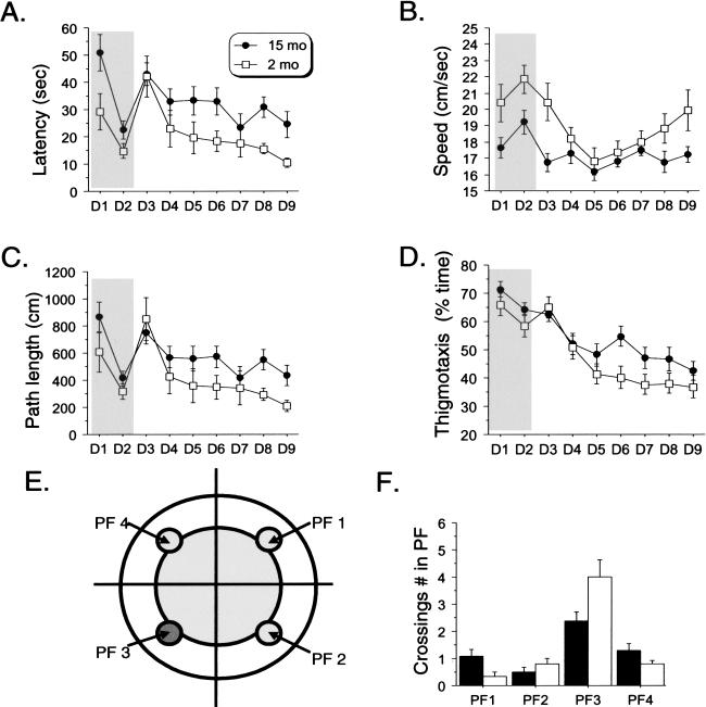
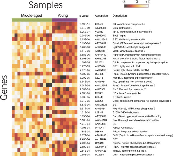
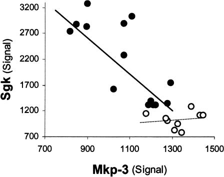
We have carried out a global survey of age-related changes in mRNA levels in the C57BL/6NIA mouse hippocampus and found a difference in the hippocampal gene expression profile between 2-month-old young mice and 15-month-old middle-aged mice correlated with an age-related cognitive deficit in hippocampal-based explicit memory formation. Middle-aged mice displayed a mild but specific deficit in spatial memory in the Morris water maze. By using Affymetrix GeneChip microarrays, we found a distinct pattern of age-related change, consisting mostly of gene overexpression in the middle-aged mice, suggesting that the induction of negative regulators in the middle-aged hippocampus could be involved in impairment of learning. Interestingly, we report changes in transcript levels for genes that could affect synaptic plasticity. Those changes could be involved in the memory deficits we observed in the 15-month-old mice. In agreement with previous reports, we also found altered expression in genes related to inflammation, protein processing, and oxidative stress.


Figures

Figure 1
Figure 1
Middle-aged mice display impaired spatial memory in the Morris Water maze. (A) Mean escape latency per day across days. Both middle-aged and young mice decreased their escape latency across both the visible (days 1 and 2) and hidden (days 3 through 9) versions of the task (significant day effect; all P < 0.001). Middle-aged mice showed lower latencies in both versions (visible: [F(1,21) = 5.00, P = -0.03]; Hidden: [F(1, 21) = 6.74, P = -0.02]). (B) Swimming speed. Significant effect of age [F(1, 21) = 7.39, P = -0.013], day [F(1, 8) = 7.28, P < 0.0001], and the interaction age × day [F(8, 168) = 2.12, P = 0.037]. (C) Path length. Both groups learned to localize the platform across both versions of the task (significant day effect: all P > 0.001). However, middle-aged mice showed impaired performance in the hidden version (days 4 through 9; significant Age effect: [F(1, 21) = 5.47, P = 0.03]), but not in the visible version (days 1 and 2 [F(1, 21) = 2.43, P = 0.13]). (D) Thigmotaxis. Significant age effect (days 4 through 9, [F(1, 21) = 5.22, P = 0.03]). (E) Scheme depicting platform location (dark grey circle in quadrant 3) and equivalent locations in the other quadrants. (F) Probe trial: crossings on previous exact spatial position of the platform (PF in quadrant 3). Middle-aged mice displayed lower performance (significant age effect: [F(1, 21) = 6.06, P = 0.02])
Figure 2
Figure 2
Age-related changes in gene expression in the mouse hippocampus. Summary of the 35 genes most significantly changed in expression between young (2 months) and middle-aged (15 months) mice. The genes are listed in order of increasing P-value. Each gene is visualized as a row of colored squares, with one square for each sample. The color indicates the relative expression of the gene according to the scale bar, with lighter colors indicating higher levels of expression. Expression levels ±2 SD from the mean of the gene are depicted as white and black, respectively. Note that the majority of genes listed showed increased expression with age. Annotations for each gene and the Welch t-test P-value (uncorrected for multiple testing) are shown at the right of the figure.
Figure 3
Figure 3
Mkp-3 and Sgk transcript levels are highly correlated in middle-aged mice in addition to showing a significant difference as compared to young mice. Sgk versus Mkp-3 hybridization signals are plotted for middle-aged (solid circles) and young (empty circles) mice. Each data point represents one chip/mouse. Sgk and Mkp-3 signals are significantly correlated within the middle-aged group (r = -0.8, P = -0.009) but not in the young animals' group (r = -0.1, P = -0.7).

References

    1. Akiyama, H., Barger, S., Barnum, S., Bradt, B., Bauer, J., Cole, G.M., Cooper, N.R., Eikelenboom, P., Emmerling, M., Fiebich, B.L., et al. 2000. Inflammation and Alzheimer's disease. Neurobiol. Aging 21: 383-421. - PMC - PubMed
    1. Amsel, A. 1993. Hippocampal function in the rat: Cognitive mapping or vicarious trial and error? Hippocampus 3: 251-256. - PubMed
    1. Armengou, A. and Davalos, A. 2002. A review of the state of research into the role of Iron in stroke. J. Nutr. Health Aging 6: 136-137. - PubMed
    1. Ashburner, M., Ball, C.A., Blake, J.A., Botstein, D., Butler, H., Cherry, J.M., Davis, A.P., Dolinski, K., Dwight, S.S., Eppig, J.T., et al. 2000. Gene ontology: Tool for the unification of biology: The gene ontology consortium. Nat. Genet. 25: 25-29. - PMC - PubMed
    1. Bach, M.E., Barad, M., Son, H., Zhuo, M., Lu, Y.F., Shih, R., Mansuy, I., Hawkins, R.D., and Kandel, E.R. 1999. Age-related defects in spatial memory are correlated with defects in the late phase of hippocampal long-term potentiation in vitro and are attenuated by drugs that enhance the cAMP signaling pathway. Proc. Natl. Acad. Sci. 96: 5280-5285. - PMC - PubMed

Publication types