. 2004 Jun;24(12):5172-83.
doi: 10.1128/MCB.24.12.5172-5183.2004.

Oxidative and osmotic stress signaling in tumor cells is mediated by ADAM proteases and heparin-binding epidermal growth factor

Affiliations

Oxidative and osmotic stress signaling in tumor cells is mediated by ADAM proteases and heparin-binding epidermal growth factor

Oliver M Fischer et al. Mol Cell Biol. 2004 Jun.

Abstract

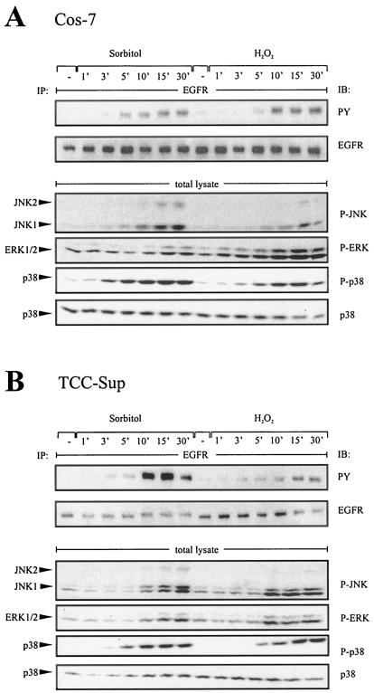
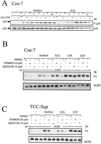
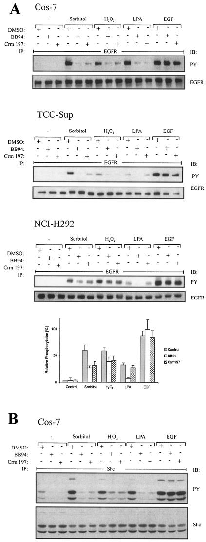
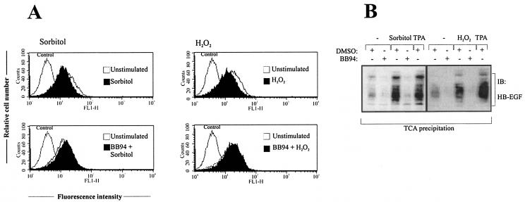
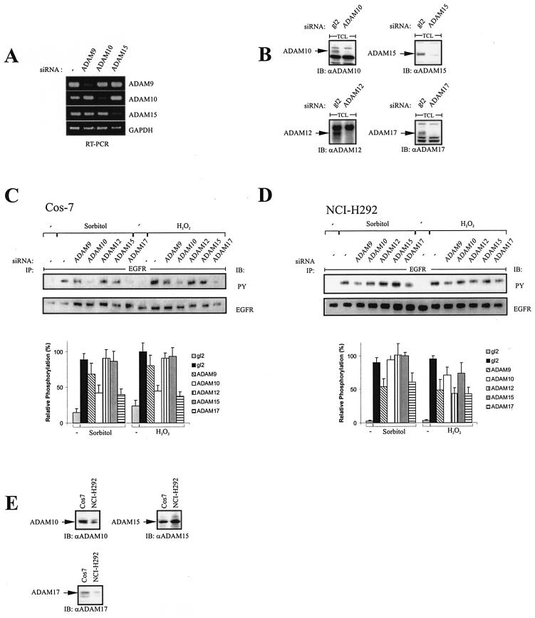
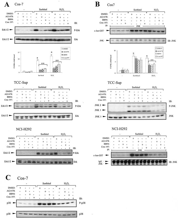
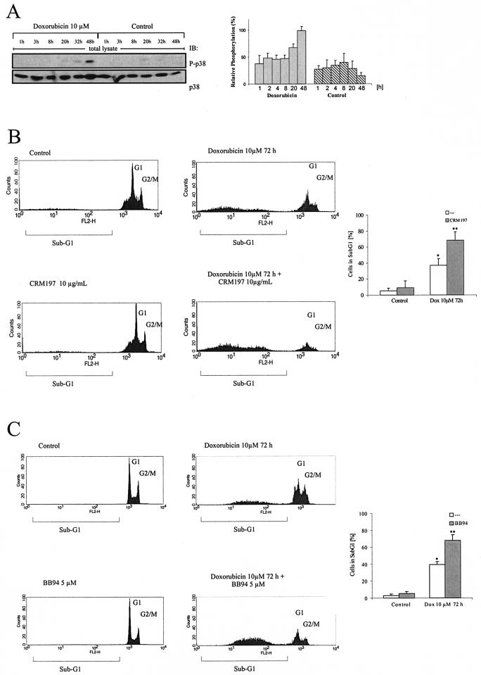
Mammalian cells respond to environmental stress by activating a variety of protein kinases critical for cellular signal transmission, such as the epidermal growth factor receptor (EGFR) tyrosine kinase and different members of the mitogen-activated protein kinase (MAPK) family. EGFR activation by stress stimuli was previously thought to occur independently of stimulation by extracellular ligands. Here, we provide evidence that osmotic and oxidative stresses induce a metalloprotease activity leading to cell surface cleavage of pro-heparin-binding EGF (pro-HB-EGF) and subsequent EGFR activation. This ligand-dependent EGFR signal resulted from stress-induced activation of the MAPK p38 in human carcinoma cells and was mediated by the metalloproteases ADAM9, -10, and -17. Furthermore, stress-induced EGFR activation induced downstream signaling through the MAPKs extracellular signal-regulated kinases 1 and 2 and JNK. Interestingly, apoptosis induced by treatment of tumor cells with doxorubicin was strongly enhanced by blocking HB-EGF function. Together, our data provide novel insights into the mammalian stress response, suggesting a broad mechanistic relevance of a p38-ADAM-HB-EGF-EGFR-dependent pathway and its potential significance for tumor cells in evasion of chemotherapeutic agent-induced apoptosis.


Figures

FIG. 1.
FIG. 1.
Time course assay of stress-induced EGFR and MAPK phosphorylation in different cell lines. (A) EGFR and MAPK phosphorylation in response to osmotic and oxidative stress. Cos-7 cells were treated with sorbitol (0.3 M) and hydrogen peroxide (200 μM) for the indicated time periods. Following immunoprecipitation (IP) of cell extracts with anti-EGFR antibody, proteins were immunoblotted (IB) with antiphosphotyrosine antibody and reprobed with anti-EGFR antibody. Phosphorylated MAPKs were detected by immunoblotting total lysates with anti-phospho-ERK, anti-phospho-JNK, and anti-phospho-p38 antibodies. The same filters were reprobed with anti-p38 antibody. (B) Stress-induced EGFR phosphorylation in human bladder carcinoma cell lines. TCC-Sup cells were treated as indicated for panel A.
FIG. 2.
FIG. 2.
p38 mediates stress-induced EGFR phosphorylation. (A) Time course assay of p38 phosphorylation dependent on EGFR kinase function. Cos-7 cells were pretreated with AG1478 (250 nM) or an equal volume of empty vehicle (dimethyl sulfoxide [DMSO]) for 20 min and stimulated with 0.3 M sorbitol or 200 μM hydrogen peroxide for the indicated periods. Cell extracts were immunoblotted with anti-phospho-p38 antibody, and the same filters were reprobed with polyclonal anti-p38 antibody. (B) Stress-induced EGFR activation depends on p38 but not ERK activity in Cos-7 cells. Cos-7 cells were pretreated with PD98059 (50 μM), SB202190 (10 μM), or an equal volume of empty vehicle (dimethyl sulfoxide) for 30 min and stimulated with 0.3 M sorbitol and 200 μM hydrogen peroxide for 10 min and the GPCR agonist LPA (10 μM) or EGF (2 ng/ml) for 3 min as a positive control. Cell extracts were assayed for EGFR tyrosine phosphorylation content. (C) Stress-induced EGFR activation depends on p38 activity in TCC-Sup carcinoma cells. TCC-Sup cells were pretreated as described for panel A and stimulated with 0.3 M sorbitol and 200 μM hydrogen peroxide for 10 min and EGF (2 ng/ml) for 3 min as a positive control. After lysis cell extracts were assayed for EGFR tyrosine phosphorylation content.
FIG.3.
FIG.3.
EGFR activation in response to osmotic and oxidative stress is ligand dependent. (A) Effect of metalloprotease and HB-EGF inhibition on EGFR phosphorylation. Cos-7, NCI-H292, and TCC-Sup cells were serum starved for 24 h; pretreated with BB94 (10 μM), the diphtheria toxin mutant Crm197 (10 μg/ml), or an equal volume of empty vehicle (dimethyl sulfoxide [DMSO]) for 20 min; and stimulated for 10 min with 0.3 M sorbitol, 200 μM hydrogen peroxide, 10 μM LPA, or 2 ng of EGF/ml. Following immunoprecipitation of cell extracts with anti-EGFR antibody, proteins were immunoblotted with antiphosphotyrosine antibody and reprobed with anti-EGFR antibody. Data from three independent experiments were quantified with the Fuji LAS1000 imaging system. (B) Analysis of Shc phosphorylation in response to stress agents. Cos-7 cells were treated as described for panel A. After immunoprecipitation of Shc from cell extracts with a polyclonal anti-Shc antibody, proteins were immunoblotted with antiphosphotyrosine antibody and reprobed with anti-Shc antibody.
FIG. 4.
FIG. 4.
Analysis of pro-HB-EGF release in response to stress agents. (A) Flow cytometric analysis of pro-HB-EGF processing. Cos-7 cells were pretreated with BB94 (10 μM) or an equal volume of empty vehicle (dimethyl sulfoxide [DMSO]) for 20 min and stimulated with 0.3 M sorbitol or 200 μM hydrogen peroxide for 30 min. Cells were collected and stained for surface pro-HB-EGF and analyzed by flow cytometry. Control cells were labeled with fluorescein isothiocyanate-conjugated secondary antibody alone. (B) Immunoblot analysis of conditioned media. Cos-7 cells were transiently transfected with pro-HB-EGF cDNA. After serum starvation for 24 h cells were stimulated for 20 min with sorbitol (0.3 M) or hydrogen peroxide (200 μM), and proteins within the supernatant medium were precipitated by TCA precipitation. Precipitated proteins were subjected to Tricine-SDS gel electrophoresis according to the protocol of Schägger and von Jargow and subsequent immunoblot analysis with anti-HB-EGF antibody. TPA stimulation has been included as a positive control. The data shown are representative of three independent experiments.
FIG. 5.
FIG. 5.
Effect of siRNAs against different ADAM proteins on stress-induced EGFR phosphorylation. (A) Blockade of ADAM metalloprotease expression by RNA interference. NCI-H292 cells were transfected with siRNA against ADAM9, ADAM10, or ADAM15; cultured for 2 days; and analyzed for gene expression by RT-PCR as indicated. GAPDH, glyceraldehyde-3-phosphate dehydrogenase. (B) Cos-7 cells were transfected with siRNAs against ADAM10, -12, -15, and -17. Forty-eight hours later, cells were lysed, protein concentration was determined, and equal amounts of protein were loaded on SDS-polyacrylamide gels. Protein levels of the respective ADAM proteases were analyzed by immunoblot analysis. (C) Cos-7 cells were transfected with siRNAs against ADAM9, -10, -12, -15, and -17; serum starved for 24 h; stimulated with 0.3 M sorbitol or 200 μM hydrogen peroxide for 10 min; and assayed for EGFR tyrosine phosphorylation content. Data from three independent experiments were quantified with the Fuji LAS1000 imaging system. (D) NCI-H292 cells were treated as described for panel C. (E) Cos-7 and NCI-H292 cells were lysed, and expression of ADAM10, -15, and -17 was assessed by immunoblot analysis as described for panel B.
FIG. 6.
FIG. 6.
MAPK activation in response to stress agents and blockade of EGFR, metalloprotease, and HB-EGF function. (A) Cos-7 cells transiently transfected with pcDNA3-HA-ERK2 and TCC-Sup and NCI-H292 cells were pretreated with AG1478 (250 nM), BB94 (10 μM), Crm197 (10 μg/ml), or an equal volume of empty vehicle (dimethyl sulfoxide [DMSO]) for 20 min and stimulated with 0.3 M sorbitol or 200 μM hydrogen peroxide for 30 min. After cell lysis total lysates were immunoblotted with anti-phospho-ERK antibody, followed by reprobing of the same membranes with polyclonal anti-ERK antibody. Quantitative analysis of ERK phosphorylation from three independent experiments (means ± standard deviations) was performed with the Fuji LAS1000 imaging system. *, P < 0.001 for control versus stimulation; **, P < 0.006 forstimulation versus stimulation plus inhibitors. (B) Cos-7 and NCI-H292 cells were treated as described for panel A. After lysis, JNK was immunoprecipitated using an anti-JNK antibody, and JNK activity was assayed using GST-c-JUN fusion protein as a substrate. Phosphorylated GST-c-JUN was visualized by autoradiography, and JNK was immunoblotted in parallel using polyclonal JNK antibody. Quantitative analysis of GST-c-JUN phosphorylation from three independent experiments (means ± standard deviations) was performed with the Fuji LAS1000 imaging system. *, P < 0.001 for control versus stimulation; **, P < 0.01 for H2O2 stimulation versus H2O2 plus inhibitors. TCC-Sup cells were treated as described for panel A. After cell lysis, JNK phosphorylation was assayed by immunoblotting cell extracts with anti-phospho-JNK antibody and the same filters were reprobed with anti-JNK antibody. (C) p38 phosphorylation is independent of EGFR, metalloprotease, and HB-EGF function. Cos-7 cells were treated as described for panel A. p38 phosphorylation was assayed by immunoblotting cell extracts with anti-phospho-p38 antibody and reprobing the same filters with anti-p38 antibody.
FIG. 7.
FIG. 7.
Doxorubicin-induced cell death of TCC-Sup carcinoma cells. (A) p38 activation in response to doxorubicin treatment. TCC-Sup cells were seeded and treated with doxorubicin for the indicated time points. After cell lysis, p38 activation was assessed by immunoblotting cell extracts with anti-phospho-p38 antibody and reprobing the same filters with anti-p38 antibody. Data from three independent experiments were quantifiedwith the Fuji LAS1000 imaging system. (B) Blockade of HB-EGF function enhances cell death in response to doxorubicin. Cells were treated for 72 h with 10 μM doxorubicin and 10 μg of Crm197/ml every 24 h as indicated. After collection of cells in assay buffer, nuclei were stained with propidium iodide and analyzed by flow cytometric analysis. Data from four independent experiments (means ± standard deviations) were quantified. *, P < 0.001 for control versus doxorubicin; **, P < 0.004 for doxorubicin versus doxorubicin plus Crm197. (C) Blockade of metalloprotease function enhances cell death in response to doxorubicin. Cells were treated and analyzed as described for panel B except that BB94 (5 μM) was used instead of Crm197. *, P < 0.003 for control versus doxorubicin; **, P < 0.007 for doxorubicin versus doxorubicin plus BB94.

References

    1. Asakura, M., M. Kitakaze, S. Takashima, Y. Liao, F. Ishikura, T. Yoshinaka, H. Ohmoto, K. Node, K. Yoshino, H. Ishiguro, H. Asanuma, S. Sanada, Y. Matsumura, H. Takeda, S. Beppu, M. Tada, M. Hori, and S. Higashiyama. 2002. Cardiac hypertrophy is inhibited by antagonism of ADAM12 processing of HB-EGF: metalloproteinase inhibitors as a new therapy. Nat. Med. 8:35-40. - PubMed
    1. Benhar, M., D. Engelberg, and A. Levitzki. 2002. ROS, stress-activated kinases and stress signaling in cancer. EMBO Rep. 3:420-425. - PMC - PubMed
    1. Biscardi, J. S., M. C. Maa, D. A. Tice, M. E. Cox, T. H. Leu, and S. J. Parsons. 1999. c-Src-mediated phosphorylation of the epidermal growth factor receptor on Tyr845 and Tyr1101 is associated with modulation of receptor function. J. Biol. Chem. 274:8335-8343. - PubMed
    1. Blanchetot, C., L. G. J. Tertoolen, and J. den Hertog. 2002. Regulation of receptor protein-tyrosine phosphatase {alpha} by oxidative stress. EMBO J. 21:493-503. - PMC - PubMed
    1. Borrell-Pages, M., F. Rojo, J. Albanell, J. Baselga, and J. Arribas. 2003. TACE is required for the activation of the EGFR by TGF-alpha in tumors. EMBO J. 22:1114-1124. - PMC - PubMed

Publication types

MeSH terms