. 2004 Jun;24(12):5404-20.
doi: 10.1128/MCB.24.12.5404-5420.2004.

RB reversibly inhibits DNA replication via two temporally distinct mechanisms

Affiliations

RB reversibly inhibits DNA replication via two temporally distinct mechanisms

Steven P Angus et al. Mol Cell Biol. 2004 Jun.

Abstract

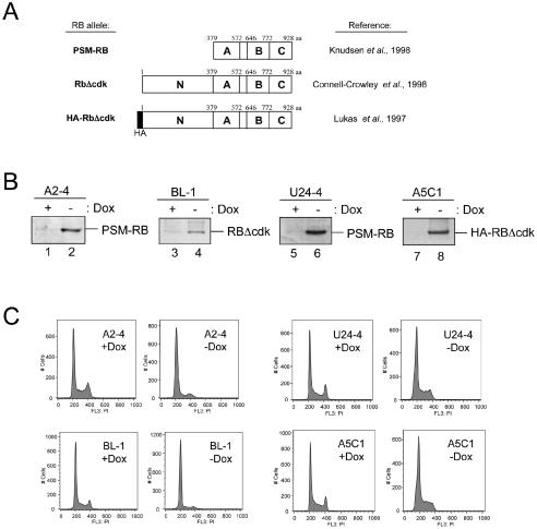
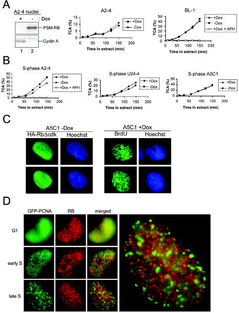
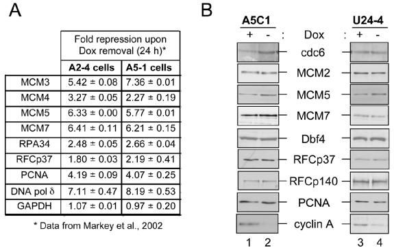
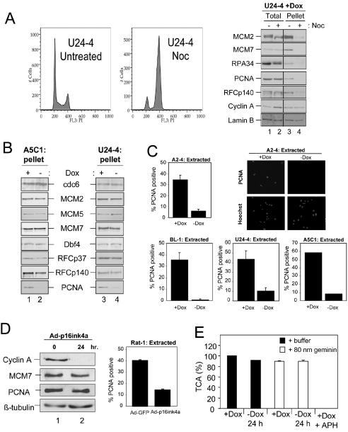
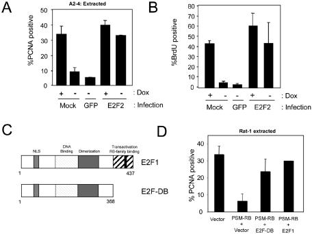
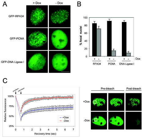
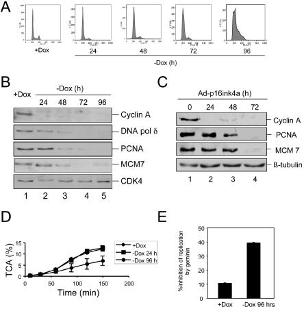
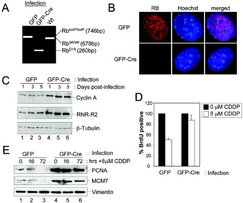
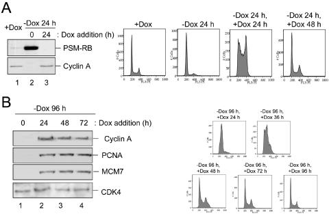
The retinoblastoma (RB) tumor suppressor is a critical negative regulator of cellular proliferation. Repression of E2F-dependent transcription has been implicated as the mechanism through which RB inhibits cell cycle progression. However, recent data have suggested that the direct interaction of RB with replication factors or sites of DNA synthesis may contribute to its ability to inhibit S phase. Here we show that RB does not exert a cis-acting effect on DNA replication. Furthermore, the localization of RB was distinct from replication foci in proliferating cells. While RB activation strongly attenuated the RNA levels of multiple replication factors, their protein expression was not diminished coincident with cell cycle arrest. During the first 24 h of RB activation, components of the prereplication complex, initiation factors, and the clamp loader complex (replication factor C) remained tethered to chromatin. In contrast, the association of PCNA and downstream components of the processive replication machinery was specifically disrupted. This signaling from RB occurred in a manner dependent on E2F-mediated transcriptional repression. Following long-term activation of RB, we observed the attenuation of multiple replication factors, the complete cessation of DNA synthesis, and impaired replicative capacity in vitro. Therefore, functional distinctions exist between the "chronic" RB-mediated arrest state and the "acute" arrest state. Strikingly, attenuation of RB activity reversed both acute and chronic replication blocks. Thus, continued RB action is required for the maintenance of two kinetically and functionally distinct modes of replication inhibition.


Figures

FIG. 1.
FIG. 1.
Cell cycle inhibition by multiple active RB alleles. (A) Diagram of active RB alles used in this study, with corresponding references. The N-terminal (N), C-terminal (C), and A and B pocket regions are identified. aa, amino acids. (B) A2-4, BL-1, U24-4, or A5C1 cell lines were cultured in the presence (+Dox) or absence (−Dox) of Dox for 24 h. Cells were harvested, and equal amounts of total protein were resolved by SDS-PAGE and immunoblotted for RB. (C) A2-4, BL-1, U24-4, or A5C1 cell lines were cultured in the presence or absence of Dox for 24 h. Cells were then fixed, stained with propidium iodide, and processed for flow cytometry. Histograms represent 10,000 gated events.
FIG. 2.
FIG. 2.
Active RB alleles do not inhibit in vitro replication or localize to replication sites. (A) The indicated cell lines were incubated in the presence (+Dox) or absence (−Dox) of Dox for 24 h. Intact nuclei were utilized for immunoblotting to detect PSM-RB and cyclin A (left panel) or resuspended in Xenopus egg extract in the presence of [α-32P]dATP or [α-32P]dCTP, and samples were removed at the indicated time points (middle and right panels). Relative DNA replication efficiency was determined by measuring the percentage of acid-precipitable cpm (TCA).(B) Nuclei from indicated cell lines were utilized as in panel A, except cultures were first synchronized in S phase by using APH. To verify the DNA polymerase-dependent incorporation of [α-32P]dATP or [α-32P]dCTP, APH was added to Xenopus egg extract as indicated. (C) A5C1 cells cultured in the presence of Dox (right panel) or in the absence of Dox for 24 h (left panel). Cells cultured in the presence of Dox were pulse-labeled with BrdU for 30 min, fixed, and stained for BrdU. Cells cultured in the absence of Dox were fixed, and immunostaining for HA-RbΔcdk was performed. (D) U2OS cells stably expressing GFP-PCNA were fixed and immunostained for endogenous RB. Representative photomicrographs of G1- and S-phase cells are shown.
FIG. 3.
FIG. 3.
RB-mediated repression of DNA replication target genes. (A) A2-4 or A5-1 cells were incubated in the presence (+Dox) or absence (−Dox) of Dox for 24 h. RNA was harvested and utilized for microarray analysis. Shown are the averages of two independent experiments comparing the relative RNA levels from the −Dox condition to the +Dox condition. These data are adapted from Markey et al. (43). (B) A5C1 and U24-4 cells were cultured in the presence (lanes 1 and 3) or absence (lanes 2 and 4) of Dox for 24 h. Equal total protein amounts were resolved by SDS-PAGE and immunoblotted for the indicated proteins.
FIG. 4.
FIG. 4.
Active RB alleles do not disrupt RFC tethering, but PCNA activity is compromised. (A) U24-4 cells were cultured either untreated or in the presence of 50-ng/ml nocodazole (Noc) for 16 h. Samples were harvested for flow cytometric analysis (left panel). Total (lanes 1 and 2) or chromatin-bound (pellet, lanes 3 and 4) fractions from untreated (lanes 1 and 3) or nocodazole-treated (lanes 2 and 4) cultures were resolved by SDS-PAGE and immunoblotted for the indicated proteins (right panel). Lamin B was utilized as a loading control. (B) A5C1 and U24-4 cells were cultured as in panel A, and equal amounts of chromatin-bound fractions (pellet) were resolved by SDS-PAGE and immunoblotted for the indicated proteins. (C) A2-4, BL-1, U24-4, and A5C1 cells were cultured in the presence (+Dox) or absence (−Dox) of Dox for 24 h. Cells were extracted in situ with CSK buffer, and PCNA was detected by immunofluorescence. Nuclei were counterstained with Hoechst stain, and the percentage of PCNA-positive cells was determined. The values shown are means ± standard deviation. At least 200 cells were counted per experiment, and representative photomicrographs are shown. (D, left panel) Asynchronously proliferating Rat-1 cells were infected with recombinant adenovirus expressing p16ink4a. Cells were harvested at 0 (lane 1) or 24 (lane 2) h postinfection. Equal total protein levels were resolved by SDS-PAGE, and the indicated proteins were detected by immunoblotting. (Right panel) Rat-1 cells were infected with GFP- or p16ink4a-encoding adenoviruses and subjected to in situ extraction with CSK buffer at 24 h postinfection. Cells were subsequently fixed, and PCNA was detected by immunofluorescence. The values shown are means ± standard deviation. At least 200 cells were counted per experiment. (E) Intact nuclei from A2-4 cells cultured in the presence or absence of Dox were resuspended in Xenopus egg extract in the presence of [α-32P]dATP that was supplemented with either buffer (filled bars) or 80 nM geminin-DEL (open bars). Following a 150-min reaction, the relative DNA replication efficiency was determined by measuring the relative percent of acid-precipitable cpm with Dox and buffer set to 100%. The values shown are means ± standard deviation.
FIG. 5.
FIG. 5.
E2F participates in the RB-mediated regulation of PCNA. (A) A2-4 cells were incubated in the presence (+Dox) or absence (−Dox) of Dox for a total of 24 h. Cells were either mock infected or infected with GFP- or E2F2-encoding adenovirus for the final 16 h. Cells were then extracted in situ and stained for PCNA. The percentage of PCNA-positive nuclei is shown as the mean ± standard deviation. More than 200 nuclei per condition were counted from two independent experiments. (B) Asynchronously proliferating A2-4 cells were cultured and infected as in panel A. BrdU incorporation was determined by indirect immunofluorescence following a 1-h pulse-labeling. The percentage of BrdU-positive nuclei is shown as the mean ± standard deviation. (C) Schematic representation of E2F1 and the E2F-DB alleles utilized. (D) Rat-1 cells were cotransfected with an H2B-GFP expression plasmid (to mark transfected cells) and the indicated plasmids. Thirty hours posttransfection, cells were subjected to in situ extraction, fixed, and stained for PCNA. The percentage of transfected (H2B-GFP positive) nuclei that were PCNA positive is shown as the mean ± standard deviation.
FIG. 6.
FIG. 6.
FRAP analysis reveals the mobilization of GFP-PCNA and downstream factors by active RB. (A) Cells harboring inducible expression of PSM-RB were transfected with GFP-RPA34, GFP-PCNA, or GFP-DNA ligase I and pBabe-puro, and stable clones were isolated by puromycin selection. Cells were synchronized in early S phase by culture in medium containing 2-μg/ml APH for 12 h. Synchronized cells were then cultured in the presence or absence of Dox for 12 h and subsequently released from APH block for 30 min prior to fixation and imaging. Bar, 5 μm. (B) The percentage of cells with focal GFP fluorescence as in panel A was determined. The values shown are means ± standard deviation. (C) The cell line stably expressing GFP-PCNA was cultured as in panel A and then utilized for FRAP analysis. Shown are the fluorescence recovery curves following 0.42 s of photobleaching (left panel) and representative images (right panel).
FIG. 7.
FIG. 7.
Chronic RB activation leads to S-phase exit. (A) A2-4 cells were cultured in the presence of Dox (+Dox) or Dox was removed and samples were collected at the indicated time points. Cells were fixed in ethanol and processed for flow cytometry. (B) Cells cultured as in panel A were harvested at the time indicated, and equal total protein was resolved by SDS-PAGE. Immunoblotting was performed to detect the indicated proteins. (C) Rat-1 cells were infected with recombinant adenovirus encoding p16ink4a, and cells were harvested at the indicated time points postinfection. Total protein was resolved by SDS-PAGE, and the indicated proteins were detected by immunoblotting. (D) A2-4 cells were cultured in the presence (+Dox) or absence (−Dox) of Dox for 96 h. Intact nuclei were prepared and incubated in Xenopus egg extract in the presence of [α-32P]dCTP. Relative DNA replication efficiency was determined by measuring the percent of acid-precipitable cpm (TCA). (E) A2-4 cells were cultured in the presence or absence of Dox for 96 h. Intact nuclei were incubated in Xenopus egg extract in the presence of [α-32P]dCTP and 80 nM geminin-DEL for 150 min. Relative DNA replication efficiency was determined by measuring the percentage of acid-precipitable cpm (TCA). The percentage of inhibition of replication by the inclusion of geminin-DEL is shown as the mean ± standard deviation.
FIG. 8.
FIG. 8.
Endogenous RB mediates both acute and chronic arrest states. (A) MAFs with a wild-type Rb gene (RbWt/Wt) or MAFs with loxP sites flanking exon 19 of the Rb gene (RbloxP/loxP) were cultured. RbloxP/loxP MAFs were infected with GFP (control)- or GFP-Cre-encoding adenovirus. Genomic DNA was isolated 72 h postinfection and utilized for PCR. Recombination/excision of exon 19 is detectable by the amplification of a novel PCR product (RbD19). (B) MAFs of the RbloxP/loxP genotype were infected with GFP- or GFP-Cre-encoding adenoviruses and cultured on glass coverslips. Cells were fixed, and endogenous RB protein was detected by immunostaining. (C) MAFs of the RbloxP/loxP genotype were infected with GFP (lanes 1 to 3)- or GFP-Cre (lanes 4 to 6)-encoding adenovirus. Cells were harvested either 1, 3, or 5 days postinfection, and equal total protein levels were resolved by SDS-PAGE and immunoblotted for the proteins indicated. (D) MAFs of the RbloxP/loxP genotype were infected with GFP- or GFP-Cre-encoding adenovirus. Seventy-two hours postinfection, cells were either untreated (0 μM) or treated with 8 μM cisplatin (CDDP) for 16 h. Subsequently the cells were pulse-labeled with BrdU, and BrdU incorporation was then monitored by immunofluorescence microscopy. The values shown are means ± standard deviation. Experiments were performed twice with greater than 200 cells counted per experiment. (E) MAFs of the RbloxP/loxP genotype infected with GFP- and Cre-encoding adenovirus were treated with 8 μM CDDP for 0 or 16 h. Cells were then cultured for an additional 56 h in drug-free medium. At the indicated times post-CDDP addition, cells were harvested and equal total protein amounts were resolved by SDS-PAGE. The indicated proteins were detected by immunoblotting.
FIG. 9.
FIG. 9.
Acute and chronic arrest states can be reversed by loss of active RB signaling. (A, left panel) A2-4 cells were cultured in the presence of Dox (+Dox, lane 1) or the absence of Dox (−Dox, lane 2). Cells were cultured in the absence of Dox for 24 h, and then Dox was added back to the media for 24 h (−Dox 24 h, +Dox 24 h, lane 3). Equal total protein amounts were resolved by SDS-PAGE, and RB and cyclin A were detected by immunoblotting. (Right panel) A2-4 cells were additionally processed as indicated for flow cytometry. (B, left panel) A2-4 cells were cultured in the absence of Dox for 96 h, and then Dox was added back to the medium for the indicated time points. Equal total protein was resolved by SDS-PAGE, and immunoblotting was performed to detect the indicated proteins. (Right panel) Samples were also utilized for flow cytometry.
FIG. 10.
FIG. 10.
RB ablation reverses the DNA damage-induced chronic arrest state. (A) Schematic diagram illustrating the protocol utilized for analysis of chronic arrest reversal in RB conditional knockout cells. (B) MAFs of the RbloxP/loxP genotype were cultured as depicted in panel A, and MCM7 was detected by immunofluorescence at the indicated time points. (Left panel) percentage of cells with MCM7-positive staining was determined. The data shown (mean ± standard deviation) are from two independent experiments with at least 150 cells counted per experiment. (Right panel) Representative photomicrographs of MCM7 immunostaining. Nuclei were counterstained with 4′,6′-diamidino-2-phenylindole (DAPI). (C) MAFs of the RbloxP/loxP genotype were cultured as depicted in panel A. Equal total protein was resolved by SDS-PAGE, and the indicated proteins were detected by immunoblotting. (D) MAFs of the RbloxP/loxP genotype were cultured as depicted in panel A. At the indicated time points, BrdU incorporation was monitored by immunofluorescent staining. The percentage of BrdU-positive cells was determined relative to untreated control. The data shown (mean ± standard deviation) are from two independent experiments with at least 150 cells counted per experiment.

References

    1. Alexander, K., and P. W. Hinds. 2001. Requirement for p27KIP1 in retinoblastoma protein-mediated senescence. Mol. Cell. Biol. 21:3616-3631. - PMC - PubMed
    1. Angus, S. P., A. F. Fribourg, M. P. Markey, S. L. Williams, H. F. Horn, J. DeGregori, T. F. Kowalik, K. Fukasawa, and E. S. Knudsen. 2002. Active RB elicits late G1/S inhibition. Exp. Cell Res. 276:201-213. - PubMed
    1. Angus, S. P., D. A. Solomon, L. Kuschel, R. F. Hennigan, and E. S. Knudsen. 2003. Retinoblastoma tumor suppressor: analyses of dynamic behavior in living cells reveal multiple modes of regulation. Mol. Cell. Biol. 23:8172-8188. - PMC - PubMed
    1. Avni, D., H. Yang, F. Martelli, F. Hofmann, W. M. ElShamy, S. Ganesan, R. Scully, and D. M. Livingston. 2003. Active localization of the retinoblastoma protein in chromatin and its response to S phase DNA damage. Mol. Cell 12:735-746. - PubMed
    1. Bartek, J., J. Bartkova, and J. Lukas. 1997. The retinoblastoma protein pathway in cell cycle control and cancer. Exp. Cell Res. 237:1-6. - PubMed

Publication types

Substances

LinkOut - more resources