. 2004 Jun;24(12):5447-58.
doi: 10.1128/MCB.24.12.5447-5458.2004.

Unconventional myosin Myo1c promotes membrane fusion in a regulated exocytic pathway

Affiliations

Unconventional myosin Myo1c promotes membrane fusion in a regulated exocytic pathway

Avirup Bose et al. Mol Cell Biol. 2004 Jun.

Abstract

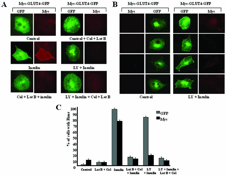
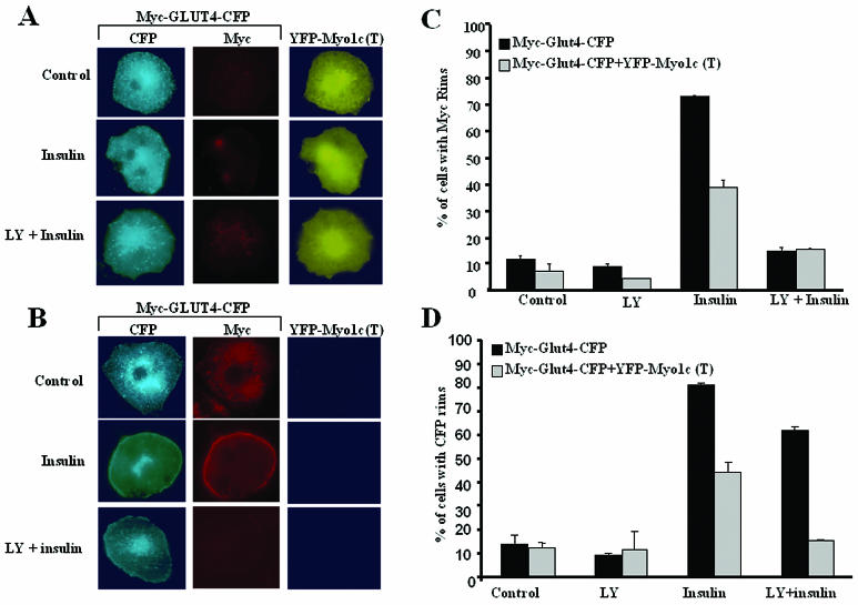
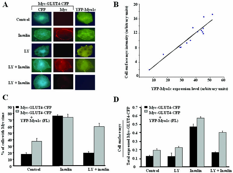
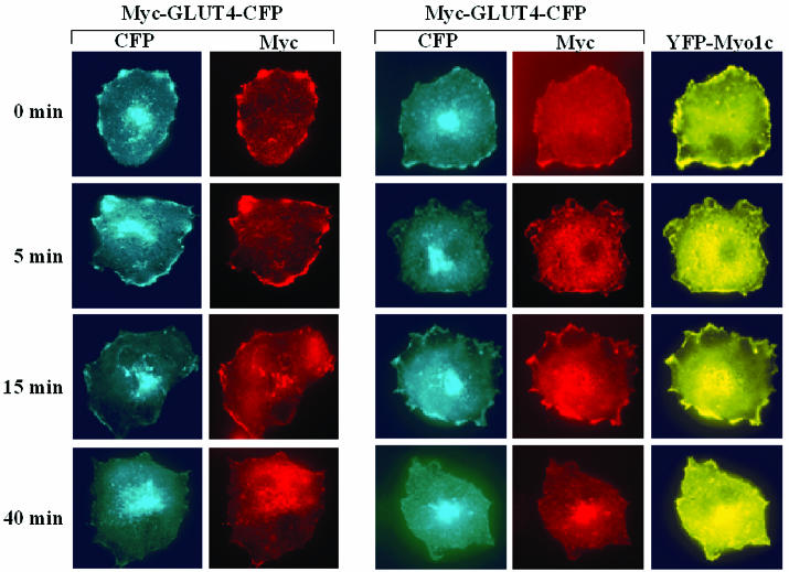
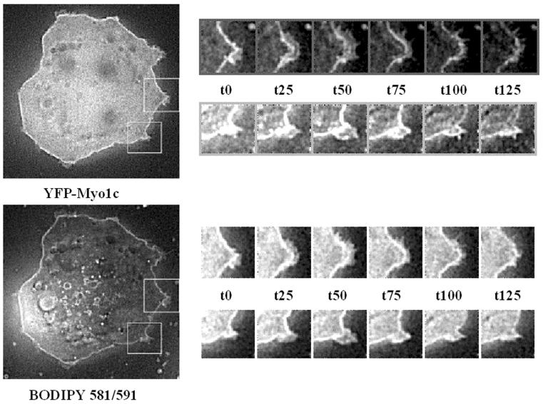
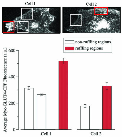
Glucose homeostasis is controlled in part by regulation of glucose uptake into muscle and adipose tissue. Intracellular membrane vesicles containing the GLUT4 glucose transporter move towards the cell cortex in response to insulin and then fuse with the plasma membrane. Here we show that the fusion step is retarded by the inhibition of phosphatidylinositol (PI) 3-kinase. Treatment of insulin-stimulated 3T3-L1 adipocytes with the PI 3-kinase inhibitor LY294002 causes the accumulation of GLUT4-containing vesicles just beneath the cell surface. This accumulation of GLUT4-containing vesicles near the plasma membrane prior to fusion requires an intact cytoskeletal network and the unconventional myosin motor Myo1c. Remarkably, enhanced Myo1c expression under these conditions causes extensive membrane ruffling and overrides the block in membrane fusion caused by LY294002, restoring the display of GLUT4 on the cell exterior. Ultrafast microscopic analysis revealed that insulin treatment leads to the mobilization of GLUT4-containing vesicles to these regions of Myo1c-induced membrane ruffles. Thus, localized membrane remodeling driven by the Myo1c motor appears to facilitate the fusion of exocytic GLUT4-containing vesicles with the adipocyte plasma membrane.


Figures

FIG. 1.
FIG. 1.
PI 3-kinase inhibitor LY294002 blocks insulin-stimulated fusion of GLUT4-containing vesicles with the plasma membrane. (A) Differentiated 3T3-L1 adipocytes expressing Myc-GLUT4-GFP were serum starved and were then either left untreated (Control), treated with colchicine and latrunculin B (Control + Col + Lat B), stimulated with insulin and treated with LY294002 followed by insulin (LY + Insulin), treated with colchicine and latrunculin B followed by insulin (Col + Lat B + insulin), or treated with LY 294002 followed by insulin for 30 min and then with colchicine and latrunculin B for 1 h (LY + Insulin + Col + Lat B). The cells were then fixed and stained with anti-Myc followed by rhodamine-labeled secondary antibody. (B) Four fields of differentiated 3T3-L1 adipocytes expressing Myc-GLUT4-GFP under serum starvation or after treatment with LY294002 (LY) and insulin from the experiment presented in panel A are shown. (C) Cells treated with the reagents mentioned above for panel A were counted for both GFP and Myc rims. At least 200 cells for each condition were counted and scored blindly for GFP and Myc rims. The images were all taken at the same exposure. The percentages of cells with GFP and Myc rims are shown. The results reflect averages for three identical experiments.
FIG. 2.
FIG. 2.
Myo1c is required to localize insulin-stimulated GLUT4-containing vesicles close to the plasma membrane. (A) Differentiated 3T3-L1 adipocytes expressing Myc-GLUT4-CFP and YFP-Myo1c(T) were serum starved and either left untreated (Control), treated with insulin (Insulin), or treated with LY294002 followed by insulin (LY + Insulin). The cells were then fixed and stained with anti-Myc followed by rhodamine-labeled secondary antibody. Cells expressing both YFP-Myo1c(T) and Myc-GLUT4-CFP are shown. (B) Cells expressing only Myc-GLUT4-CFP. (C) Cells from panels A and B were counted for Myc rims at the cell surfaces. (D) Cells from panels A and B were counted for CFP signals at the cell surfaces. More than 100 cells were scored blindly for Myc and CFP rims. The data represent averages for three similar experiments.
FIG. 3.
FIG. 3.
Myo1c expression partially overcomes the LY294002-induced block in fusion of GLUT4-containing vesicles. (A) Differentiated 3T3-L1 adipocytes expressing Myc-GLUT4-CFP and YFP-Myo1c were serum starved and then either left untreated (Control), treated with insulin (Insulin), or treated with LY294002 followed by insulin (LY + Insulin). The top four rows of panels show cells expressing both Myc-GLUT4-CFP and YFP-Myo1c, and the fifth row of panels show a cell expressing only Myc-GLUT4-CFP. (B) Differentiated 3T3-L1 adipocytes expressing Myc-GLUT4-CFP and YFP-Myo1c were stimulated with insulin. The cell surface Myc signal (rhodamine) intensity in these cells was quantitated and compared with the YFP-Myo1c signal intensity in these cells. (C) Cells shown in panel A were counted for anti-Myc rims at the cell surfaces. More than 100 cells for each condition were scored blindly for Myc rims. (D) Quantification of cell surface anti-Myc signal (rhodamine) intensity in the cells shown in panel A. The arbitrary unit represents the ratio of the cell surface anti-Myc signal to the total CFP signal in each cell. The data represent averages for five similar experiments.
FIG. 4.
FIG. 4.
Myo1c expression does not disrupt Myc-GLUT4-CFP internalization to the perinuclear regions of 3T3-L1 adipocytes. Differentiated 3T3-L1 adipocytes either transfected with Myc-GLUT4-CFP alone or cotransfected with Myc-GLUT4-CFP and YFP-Myo1c were stimulated with insulin for 30 min. The cells were then labeled with anti-Myc antibodies and then warmed to 37°C for the indicated times to allow the Myc-GLUT4-CFP to undergo endocytosis. The cells were then permeabilized and then stained with rhodamine-conjugated secondary antibody to detect total Myc-GLUT4-CFP. The data are representative of three similar experiments.
FIG. 5.
FIG. 5.
Myo1c expression in 3T3-L1 adipocytes causes insulin-independent membrane ruffles. Differentiated 3T3-L1 adipocytes expressing YFP-Myo1c were serum starved. BODIPY 581/591 was added to the media at a final concentration of 1 μM, and the cells were incubated for 20 min at 37°C. Membrane ruffling was observed by monitoring the cells live for 10 min, imaging them at 5-s intervals. Shown are six frames, each 25 s apart, for both BODIPY 581/591 stain and YFP-Myo1c. The images are all single optical sections from three-dimensional images following image restoration.
FIG. 6.
FIG. 6.
Ultrafast microscopy reveals enrichment of GLUT4 and Myo1c in ruffling areas compared to nonruffling areas. (Top) Three-dimensional projection images of two different cells, showing Myc-GLUT4-CFP distribution. The images shown were selected from a sequence of 100 such three-dimensional images of each cell spanning 16.7 min (see Materials and Methods). The three-dimensional images were first subjected to image restoration to remove out-of-focus light, and then maximum-intensity projections were made by retaining the maximum (brightest) intensity at each pixel position of the 21 optical sections. Regions of active membrane ruffling (red boxes) were identified visually and compared with regions not showing significant ruffling (white boxes). See the supplemental material for complete movie sequences. (Bottom) In order to separate cytosolic from membrane-associated Myc-GLUT4-CFP fluorescence, an intensity threshold was chosen for each cell such that 95% of the Myc-GLUT4-CFP pixels in the cytosol were below the threshold. Then, for each region of each cell, the average Myc-GLUT4-CFP fluorescence of just the pixels whose intensities were above the threshold and within the indicated region was computed for each time point. The mean and standard deviation of the average Myc-GLUT4-CFP concentration for all 100 time points were calculated and plotted as shown.

References

    1. Bennett, M. K., J. E. Garcia-Arraras, L. A. Elferink, K. Peterson, A. M. Fleming, C. D. Hazuka, and R. H. Scheller. 1993. The syntaxin family of vesicular transport receptors. Cell 74:863-873. - PubMed
    1. Borisy, G. G., and T. M. Svitkina. 2000. Actin machinery: pushing the envelope. Curr. Opin. Cell Biol. 12:104-112. - PubMed
    1. Bose, A., A. Guilherme, S. I. Robida, S. M. C. Nicoloro, Q. L. Zhou, Z. Y. Jiang, D. P. Pomerleau, and M. P. Czech. 2002. Glucose transporter recycling in response to insulin is facilitated by myosin Myo1c. Nature 420:821-824. - PubMed
    1. Bretscher, M. S., and C. Aguado-Velasco. 1998. EGF induces recycling membrane to form ruffles. Curr. Biol. 8:721-724. - PubMed
    1. Broadie, K., A. Prokop, H. J. Bellen, C. J. O'Kane, K. L. Schulze, and S. T. Sweeney. 1995. Syntaxin and synaptobrevin function downstream of vesicle docking in Drosophila. Neuron 15:663-673. - PubMed

Publication types

MeSH terms