Endothelin-induced constriction of the ductus venosus in fetal sheep: developmental aspects and possible interaction with vasodilatory prostaglandin
- PMID: 15172962
- PMCID: PMC1575056
- DOI: 10.1038/sj.bjp.0705849
Endothelin-induced constriction of the ductus venosus in fetal sheep: developmental aspects and possible interaction with vasodilatory prostaglandin
Abstract
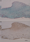
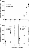
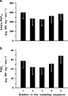
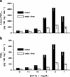
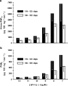
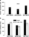
1. The ductus venosus is actively regulated in the fetus, but questions remain on the presence of a functional sphincter at its inlet. Using fetal sheep (0.6-0.7 gestation onwards), we have examined the morphology of the vessel and have also determined whether endothelin-1 (ET-1) qualifies as a natural constrictor being modulated by prostaglandins (PGs). 2. Masson's staining and alpha-actin immunohistochemistry showed a muscular, sphincter-like formation at the ductus inlet and a muscle layer within the wall of the vessel proper. This muscle cell component increased with age. 3. ET-1 contracted dose-dependently isolated sphincter and extrasphincter preparations of the ductus from term fetus. This ET-1 effect also occurred in the premature, but its threshold was higher. 4. BQ123 (1 microm) caused a rightward shift in the ET-1 dose-response curve, while indomethacin at a threshold concentration (28 nm) tended to have an opposite effect. 5. Big ET-1 also contracted the ductus sphincter but differed from ET-1 for its lesser potency and inhibition by phosphoramidon (50 microm). 6. The ductus sphincter (term and preterm) and extrasphincter (term) released 6-keto-PGF(1alpha) (hence PGI(2)) and, to a lesser degree, PGE(2) at rest and their release increased dose-dependently upon ET-1 treatment. Both basal and stimulated release was curtailed by endothelium removal. 7. BQ123 and phosphoramidon reduced slightly the contraction of ductus sphincter to indomethacin (2.8 microm). 8. We conclude that the ductus contains a contractile mechanism in the sphincter and extrasphincter regions. ET-1 lends itself to a role in the generation of contractile tone and its action may be modulated by prostaglandins.
Figures



Similar articles
-
Involvement of endothelin-1 in hypoxic pulmonary vasoconstriction in the lamb.J Physiol. 1995 Jan 15;482 ( Pt 2)(Pt 2):421-34. doi: 10.1113/jphysiol.1995.sp020529. J Physiol. 1995. PMID: 7714833 Free PMC article.
-
Evidence for a role of prostaglandin I2 and thromboxane A2 in the ductus venosus of the lamb.Can J Physiol Pharmacol. 1985 Sep;63(9):1101-5. doi: 10.1139/y85-181. Can J Physiol Pharmacol. 1985. PMID: 3902179
-
The response of the lamb ductus arteriosus to endothelin: developmental changes and influence of light.Life Sci. 2002 Jul 26;71(10):1209-17. doi: 10.1016/s0024-3205(02)01822-2. Life Sci. 2002. PMID: 12095541
-
The control of the ductus venosus: an update.Eur J Pediatr. 1993 Dec;152(12):976-7. doi: 10.1007/BF01957218. Eur J Pediatr. 1993. PMID: 8131814 Review. No abstract available.
-
The control of cardiovascular shunts in the fetal and perinatal period.Can J Physiol Pharmacol. 1988 Aug;66(8):1129-34. doi: 10.1139/y88-185. Can J Physiol Pharmacol. 1988. PMID: 3052747 Review.
Cited by
-
Altered subcellular localization of heat shock protein 90 is associated with impaired expression of the aryl hydrocarbon receptor pathway in dogs.PLoS One. 2013;8(3):e57973. doi: 10.1371/journal.pone.0057973. Epub 2013 Mar 5. PLoS One. 2013. PMID: 23472125 Free PMC article.
References
-
- ADEAGBO A.S.O., BISHAI I., LEES J., OLLEY P.M., COCEANI F. Evidence for a role of prostaglandin I2 and thromboxane A2 in the ductus venosus of the lamb. Can. J. Physiol. Pharmacol. 1985;63:1101–1105. - PubMed
-
- ADEAGBO A.S.O., BREEN C.A., CUTZ E., LEES J.G., OLLEY P.M., COCEANI F. Lamb ductus venosus: evidence of a cytochrome P-450 mechanism in its contractile tension. J. Pharmacol. Exp. Ther. 1990;252:875–879. - PubMed
-
- ADEAGBO A.S.O., COCEANI F., OLLEY P.M. The response of the lamb ductus venosus to prostaglandin and inhibitors of prostaglandin and thromboxane synthesis. Circ. Res. 1982;51:580–586. - PubMed
-
- AILAMAZYAN E.K., KIRILLOVA O.V., POLYANIN A.A., KOGAN I.Y. Functional morphology of ductus venosus in human fetus. Neuroendocrinol. Lett. 2003;24:28–32. - PubMed
-
- BELLOTTI M., PENNATI G., DE GASPERI C., BATTAGLIA F.C., FERRAZZI E. Role of ductus venosus in distribution of umbilical blood flow in human fetuses during second half of pregnancy. Am. J. Physiol. 2000;279:H1256–H1263. - PubMed
Publication types
MeSH terms
Substances
LinkOut - more resources
Full Text Sources

