. 2004 Jun 7;165(5):735-46.
doi: 10.1083/jcb.200312170. Epub 2004 Jun 1.

Dynamics of putative raft-associated proteins at the cell surface

Affiliations

Dynamics of putative raft-associated proteins at the cell surface

Anne K Kenworthy et al. J Cell Biol. .

Abstract

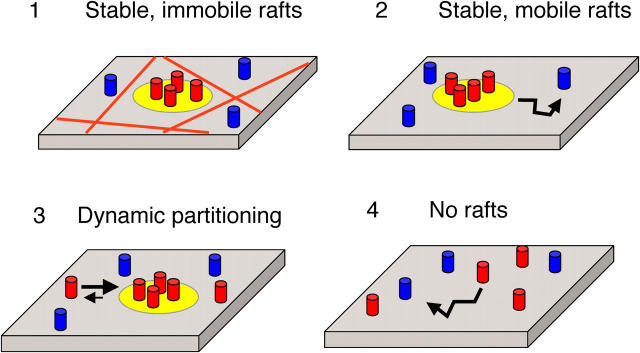
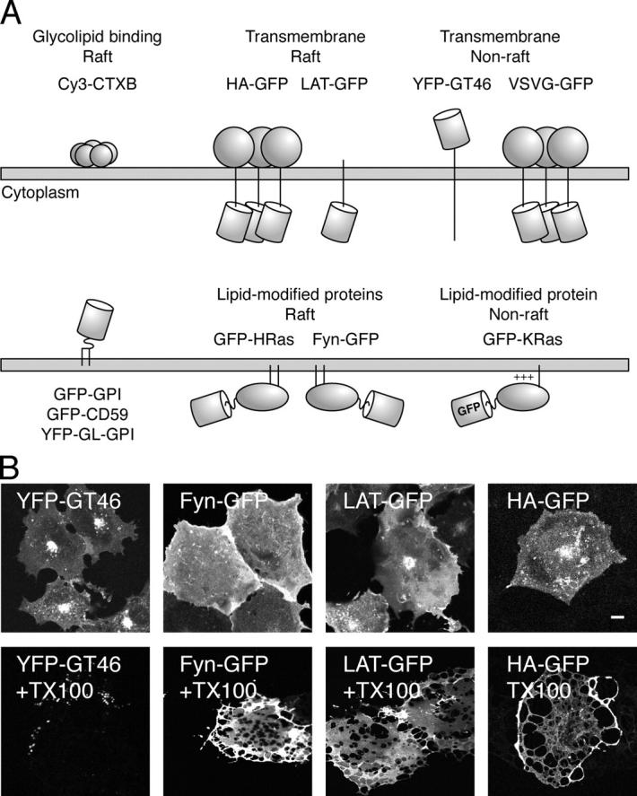
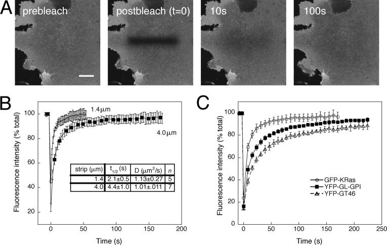
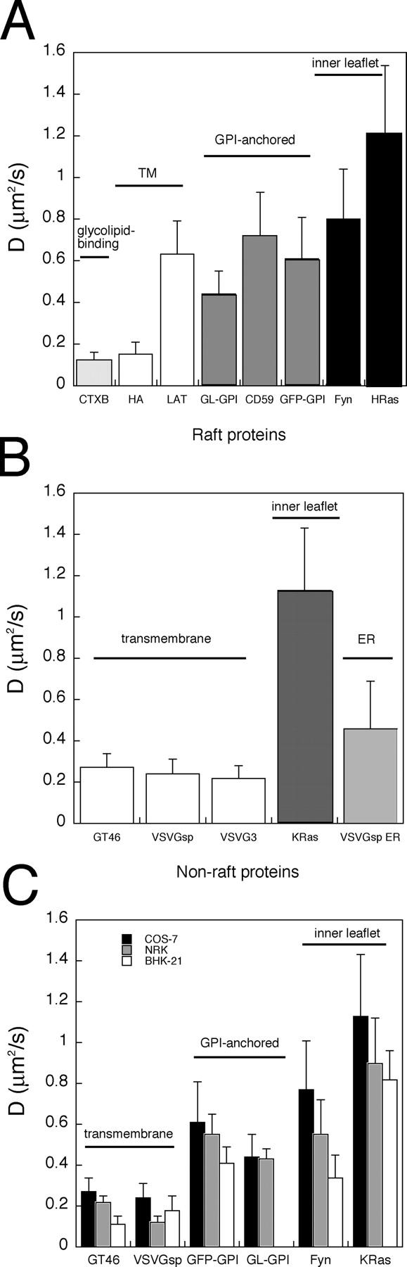
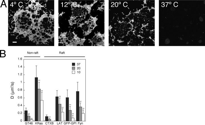
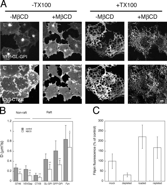
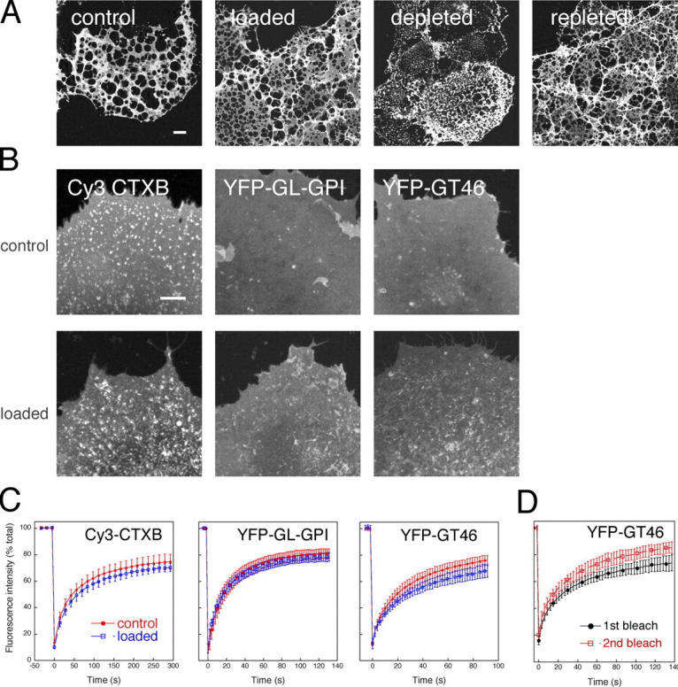
Lipid rafts are conceptualized as membrane microdomains enriched in cholesterol and glycosphingolipid that serve as platforms for protein segregation and signaling. The properties of these domains in vivo are unclear. Here, we use fluorescence recovery after photobleaching to test if raft association affects a protein's ability to laterally diffuse large distances across the cell surface. The diffusion coefficients (D) of several types of putative raft and nonraft proteins were systematically measured under steady-state conditions and in response to raft perturbations. Raft proteins diffused freely over large distances (> 4 microm), exhibiting Ds that varied 10-fold. This finding indicates that raft proteins do not undergo long-range diffusion as part of discrete, stable raft domains. Perturbations reported to affect lipid rafts in model membrane systems or by biochemical fractionation (cholesterol depletion, decreased temperature, and cholesterol loading) had similar effects on the diffusional mobility of raft and nonraft proteins. Thus, raft association is not the dominant factor in determining long-range protein mobility at the cell surface.


Figures

Figure 1.
Figure 1.
Models for lipid raft dynamics and protein diffusional mobility. Models for diffusional mobility of lipid rafts (yellow), raft-associated proteins (red), and nonraft proteins (blue). (1) Stable, immobile rafts. Hypothetical barriers to lipid raft diffusion are depicted by the red lines. (2) Stable, mobile rafts. (3) Dynamic partitioning of raft proteins. (4) No rafts. For simplicity, putative barriers to individual protein diffusion are not depicted. See text for further details.
Figure 2.
Figure 2.
Proteins used in this work. (A) Membrane topology of GFP/YFP chimeras and fluorescently labeled toxin used in this work. The barrel indicates the position of the GFP. (B) Extraction of COS-7 cells grown on coverslips with 1% TX-100 at 4°C confirms that Fyn-GFP, LAT-GFP, and HA-GFP are present in DRM. YFP-GT46, a nonraft protein, is essentially completely solubilized under identical conditions. Bar, 10 μm.
Figure 3.
Figure 3.
Large-scale lateral diffusion measurements by confocal microscopy. (A) Selected images from a confocal FRAP experiment at 37°C of GFP-KRas expressed in COS-7 cells. Bleach box, 4 μm wide. Bar, 10 μm. (B) Kinetics of recovery for 1.4- (circles) versus 4-μm-wide (squares) bleach box. Calculated D and t1/2 values are indicated. Data shown are for GFP-KRas expressed in COS-7 cells at 37°C. (C) Kinetics of recovery for YFP-GT46 (triangles), YFP-GL-GPI (squares), and GFP-KRas (circles) in COS-7 cells at 37°C using a 4-μm-wide bleach box. Each curve shows the mean ± SD from seven to nine cells from a single experiment. The calculated Ds were as follows: GFP-KRas, 1.01 ± 0.11 μm2/s; YFP-GL-GPI, 0.47 ± 0.07 μm2/s; YFP-GT46, 0.23 ± 0.02 μm2/s.
Figure 4.
Figure 4.
Diffusional mobilities of raft and nonraft proteins at the cell surface at 37°C. D for raft proteins (A) and for nonraft proteins (B) in COS-7 cells. (C) D for a subset of proteins in COS-7, BHK-21, and NRK cells. The bleach box was 4 μm wide in COS-7 cells and 1.4 μm wide in NRK and BHK-21 cells. Error bars are the mean ± SD.
Figure 5.
Figure 5.
Effect of temperature on detergent solubility and lateral mobility of raft and nonraft proteins. (A) The fraction of DRM decreases with increasing temperature of extraction in 1% TX-100. DRM was visualized by Cy3-CTXB labeling. Bar, 10 μm. (B) D decreases with decreasing temperature for both raft and nonraft proteins. **, P < 0.0001, t test. Error bars are the mean ± SD.
Figure 6.
Figure 6.
Effect of cholesterol depletion on detergent solubility and lateral mobility of raft and nonraft proteins, and quantitation of filipin staining after perturbations in cellular cholesterol levels. (A) Cholesterol depletion enhances the TX-100 solubility of YFP-GL-GPI and Cy3-CTXB. A similar effect was observed for other raft proteins (not depicted). Bar, 10 μm. (B) Cholesterol depletion slows the diffusion of raft and nonraft proteins at 37°C. Cells were pretreated with 10 mM MβCD for 30 min at 37°C before TX-100 extraction or FRAP measurements. **, P < 0.0001; *, P < 0.01, t test. Error bars are the mean ± SD. (C) Quantitation of cholesterol by filipin staining. This method, although not strictly quantitative (Severs, 1997; Maxfield and Wüstner, 2002), provides a qualitative estimate of the cholesterol levels. Filipin staining was performed on fixed cells and quantified from images obtained using a CCD camera for identical exposure times as described in Materials and methods. Data are shown from a representative experiment. Error bars are the mean ± SD. All treatments are significantly different than controls (P < 0.0001, t test).
Figure 7.
Figure 7.
Effect of cholesterol loading on detergent solubility, cell surface distribution, and lateral mobility of raft and nonraft proteins. (A) Comparison of DRM in mock-treated, cholesterol-loaded, cholesterol-depleted, and cholesterol-repleted cells. DRM were visualized by Cy3-CTXB labeling. Bar, 10 μm. (B) Effect of cholesterol loading on the cell surface distribution of raft (Cy3-CTXB and YFP-GL-GPI) and nonraft (YFP-GT46) proteins in living cells. Averaged prebleach images from confocal FRAP experiments are shown for control and cholesterol-loaded cells. Bar, 10 μm. (C) Effect of cholesterol loading on diffusional mobilities of raft proteins and nonraft proteins. Recovery curves show the mean ± SD from 7–11 cells from a representative experiment for control (red circles) versus cholesterol-loaded (blue squares) cells at 22°C. (D) Repetitive bleaching of YFP-GT46 in cholesterol-loaded cells to test for the presence of an immobile pool of protein. After performing a FRAP measurement on a given cell (first bleach, black circles), a second measurement was made using exactly the same bleach box (second bleach, red squares). Recovery curves show the mean ± SD from 7–11 cells from a representative experiment at 22°C.

References

    1. Anderson, R.G.W., and K. Jacobson. 2002. A role for lipid shells in targeting proteins to caveolae, rafts and other lipid domains. Science. 296:1821–1825. - PubMed
    1. Aroeti, B., and Y.I. Henis. 1988. Effects of fusion temperature on the lateral mobility of Sendai virus glycoproteins in erythrocyte membranes and on cell fusion indicate that glycoprotein mobilization is required for cell fusion. Biochemistry. 27:5654–5661. - PubMed
    1. Bacia, K., I.V. Majoul, and P. Schwille. 2002. Probing the endocytic pathway in live cells using dual-color fluorescence cross-correlation analysis. Biophys. J. 83:1184–1193. - PMC - PubMed
    1. Brown, D.A., and J.K. Rose. 1992. Sorting of GPI-anchored proteins to glycolipid-enriched membrane subdomains during transport to the apical cell surface. Cell. 68:533–544. - PubMed
    1. Brown, D.A., and E. London. 1998. a. Functions of lipid rafts in biological membranes. Annu. Rev. Cell Dev. Biol. 14:111–136. - PubMed

Publication types