. 2004 Jun 7;165(5):653-62.
doi: 10.1083/jcb.200309097. Epub 2004 Jun 1.

Ionophore-resistant mutant of Toxoplasma gondii reveals involvement of a sodium/hydrogen exchanger in calcium regulation

Affiliations

Ionophore-resistant mutant of Toxoplasma gondii reveals involvement of a sodium/hydrogen exchanger in calcium regulation

Gustavo Arrizabalaga et al. J Cell Biol. .

Abstract

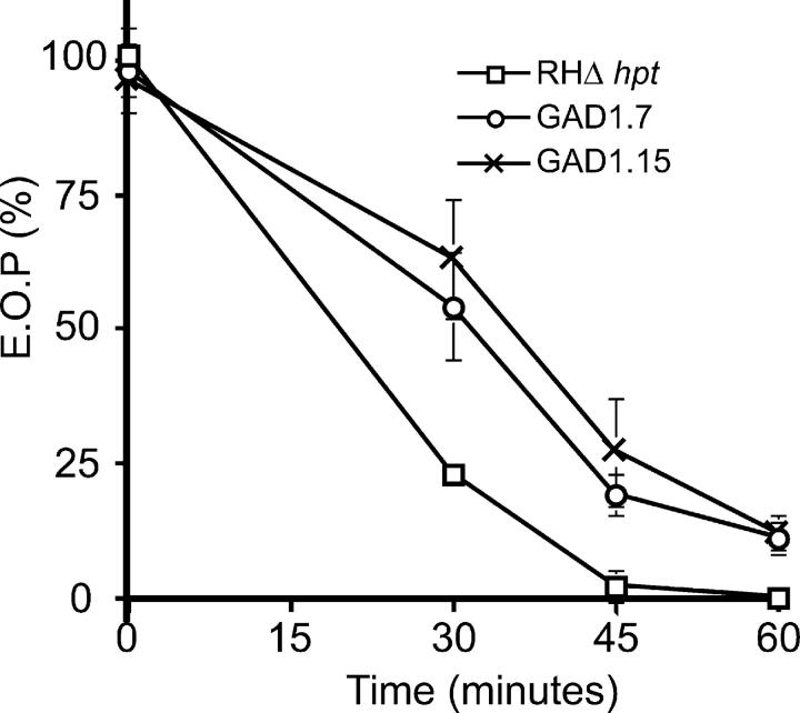
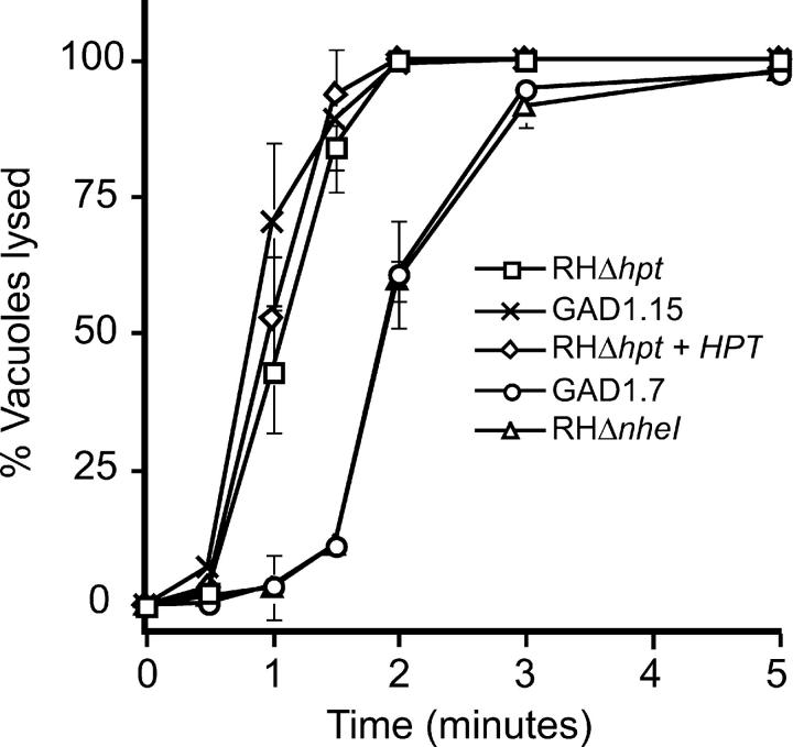
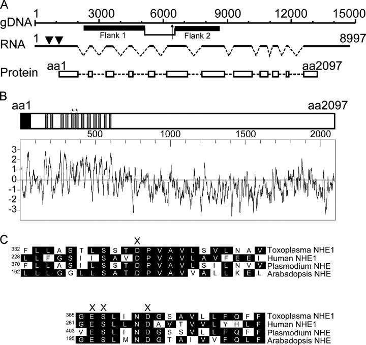
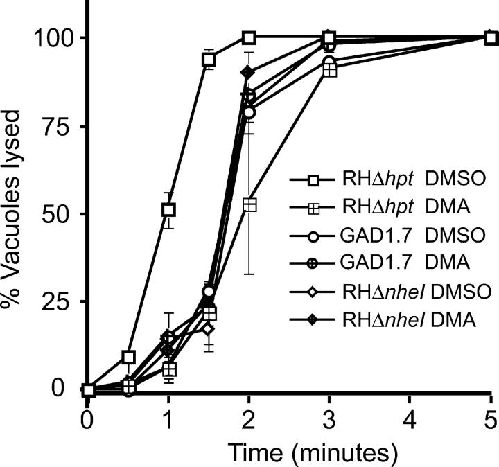
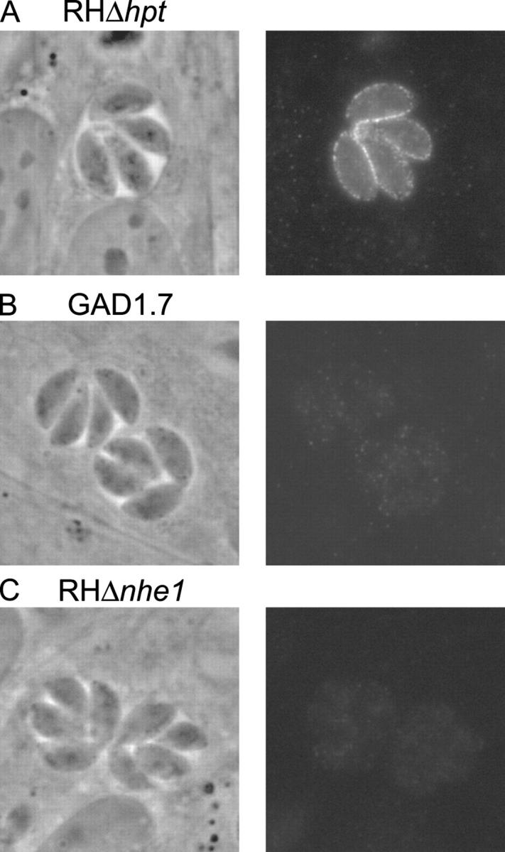
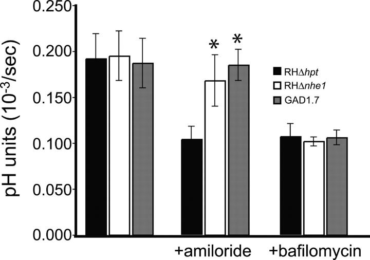
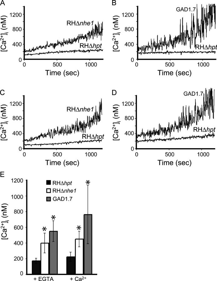
Calcium is a critical mediator of many intracellular processes in eukaryotic cells. In the obligate intracellular parasite Toxoplasma gondii, for example, a rise in [Ca2+] is associated with significant morphological changes and rapid egress from host cells. To understand the mechanisms behind such dramatic effects, we isolated a mutant that is altered in its responses to the Ca2+ ionophore A23187 and found the affected gene encodes a homologue of Na+/H+ exchangers (NHEs) located on the parasite's plasma membrane. We show that in the absence of TgNHE1, Toxoplasma is resistant to ionophore-induced egress and extracellular death and amiloride-induced proton efflux inhibition. In addition, the mutant has increased levels of intracellular Ca2+, which explains its decreased sensitivity to A23187. These results provide direct genetic evidence of a role for NHE1 in Ca2+ homeostasis and important insight into how this ubiquitous pathogen senses and responds to changes in its environment.


Figures

Figure 1.
Figure 1.
IID phenotype of the parental strain RHΔhpt and two Iid mutants, GAD1.7 and GAD1.15. Extracellular parasites were exposed to A23187 and then assayed for the ability to form plaques. Efficiency of plating (EOP) was defined as the percentage of plaque-forming units arising from parasites incubated with A23187 versus the DMSO-treated control. Each data point represents the average of three experiments and the error bars represent the standard deviation.
Figure 2.
Figure 2.
IIE phenotype of GAD and knockout mutants. Percentage of vacuoles lysed at specific time points after induction with 1 μM A23187 in DMEi was measured for the parental strain RHΔhpt, the two Iid mutants GAD1.7 and GAD1.15, the NHE1 knockout strain RHΔnhe1, and the HXGPRT-expressing strain RHΔhpt+HPT. Each data point represents the average of three experiments, and the error bars represent the standard deviation.
Figure 3.
Figure 3.
Genomic arrangement and protein domains of NHE1 . The Toxoplasma NHE1 gene encodes a protein 2,097 amino acids long with 12 TM domains. (A) The distribution of exons (dark lines), introns (broken lines), and coding sequences (white boxes) along the genomic region encoding NHE1 are shown. Base 1 in the gDNA and RNA graph represents the transcription start site as determined by 5′ RACE (nucleotide 19397 in TGG_10551, ToxoDB v.2.1). Also shown are the genomic regions (black boxes) used as flanking fragments in the generation of the NHE1 knockout construct. The bracket in the gDNA drawing indicates the region that is deleted and replaced by the HPT gene in the RHΔnhe1 knockout strain, while the arrow shows the site of the original insertion in the GAD1.7 mutant. The arrowheads in the RNA indicate the position of the two putative translation initiation sites. (B) Shown is the schematic of the TgNHE1 protein, with its signal sequence (black box) and the predicted 12 TM domains (gray boxes). Below and in the same scale is the Kyte-Doolitle hydropathy plot for the entire protein (Kyte and Doolittle, 1982). (C) Sequence comparison of the two highly conserved TM domains in Toxoplasma (NHE1, TM domains indicated by * in B), human (NHE1) (Sardet et al., 1988), P. falciparum (putative NHE) (PlasmoDB.org), and Arabadopsis (NHE/SOS1) (Shi et al., 2000) is shown. The conserved polar amino acids proposed to be involved in cation binding and translocation are marked by X (Wiebe et al., 2001). The amino acid number of the first residue in each line is shown. Complete sequence of TgNHE1 is available from GenBank/EMBL/DDBJ, accession no. AY485268.
Figure 4.
Figure 4.
Effect of pretreating parasites with an NHE inhibitor on IIE. Percentage of vacuoles lysed at specific time points after induction with 1 μM A23187 in DMEi was measured for RHΔhpt, GAD1.7, and RHΔnhe1 parasites that were pretreated with either 100 μM DMA dissolved in DMSO or an equivalent amount of DMSO alone for 10 min. The data are pooled from three independent trials, and the error bars represent the standard deviation.
Figure 5.
Figure 5.
Intracellular localization of TgNHE1. Intracellular parasites of either (A) the wild-type RHΔhpt, (B) the mutant GAD1.7, or (C) the NHE knockout RHΔnhe1 were stained with antibodies raised in mouse against amino acids 716–1073 of the TgNHE1 protein. An Alexa fluor 488–conjugated goat anti–mouse antibody was used to visualize the TgNHE1 signal. The phase contrast and fluorescence images of the same vacuole are shown for each strain.
Figure 6.
Figure 6.
Effect of NHE inhibition on proton efflux. The level of proton efflux was measured for wild-type parasites (RHΔhpt) or either of the NHE1 mutants GAD1.7 and RHΔnhe1 by incubating the parasites in a weakly buffered solution in the presence of BCECF. The effect of the NHE inhibitor amiloride and the V-H+-ATPase inhibitor bafilomycin A1 on proton efflux for each strain is shown. Each bar represents the average of six independent experiments, and the error bars represent the standard deviation. Values that are statistically different from the RHΔhpt control as assessed by a t test are marked by * (P < 0.05).
Figure 7.
Figure 7.
Ca 2+ homeostasis in the parental strain RHΔhpt and the nhe mutants RHΔnhe1 and GAD1.7. Intracellular Ca2+ concentration of Fura-2AM–loaded parasites was measured in the presence of either EGTA (A and B) or CaCl2 (C and D) by loading the parasites with Fura-2AM. Traces shown are from representative experiments. (E) The average [Ca2+]i for each of the three strains at 10 min in the presence of either EGTA or CaCl2 is shown. Each bar represents the average of at least three independent experiments, and the error bars represent the standard deviation. Values that are statistically different from the RHΔhpt control as assessed by a t test are marked by * (P < 0.015).

Similar articles

Cited by

References

    1. Berthe, P., J.L. Cousin, and J.P. Breittmayer. 1991. Intracellular Ca2+ regulation in CD3 stimulated Jurkat T cells involves H+ fluxes. Cell. Signal. 3:453–459. - PubMed
    1. Black, M.W., and J.C. Boothroyd. 1998. Development of a stable episomal shuttle vector for Toxoplasma gondii. J. Biol. Chem. 273:3972–3979. - PubMed
    1. Black, M.W., and J.C. Boothroyd. 2000. Lytic cycle of Toxoplasma gondii. Microbiol. Mol. Biol. Rev. 64:607–623. - PMC - PubMed
    1. Black, M.W., G. Arrizabalaga, and J.C. Boothroyd. 2000. Ionophore-resistant mutants of Toxoplasma gondii reveal host cell permeabilization as an early event in egress. Mol. Cell. Biol. 20:9399–9408. - PMC - PubMed
    1. Bosia, A., D. Ghigo, F. Turrini, E. Nissani, G.P. Pescarmona, and H. Ginsburg. 1993. Kinetic characterization of Na+/H+ antiport of Plasmodium falciparum membrane. J. Cell. Physiol. 154:527–534. - PubMed

Publication types

MeSH terms