. 2004 Jun 7;199(11):1577-84.
doi: 10.1084/jem.20040168. Epub 2004 Jun 1.

Short-lived plasmablasts and long-lived plasma cells contribute to chronic humoral autoimmunity in NZB/W mice

Affiliations

Short-lived plasmablasts and long-lived plasma cells contribute to chronic humoral autoimmunity in NZB/W mice

Bimba F Hoyer et al. J Exp Med. .

Abstract

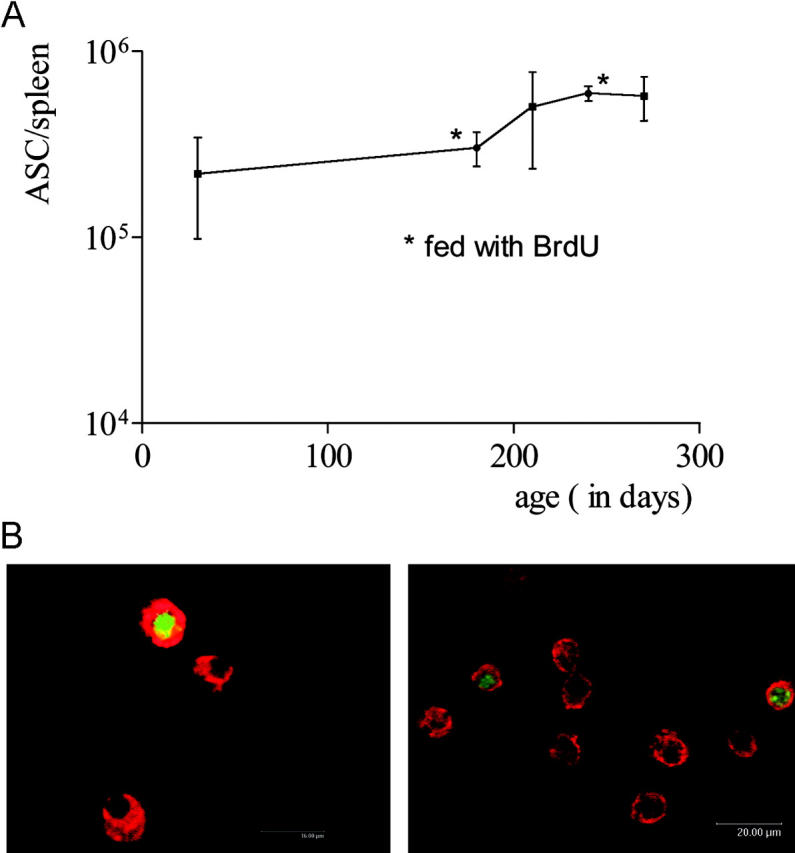
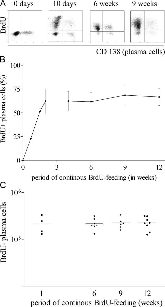
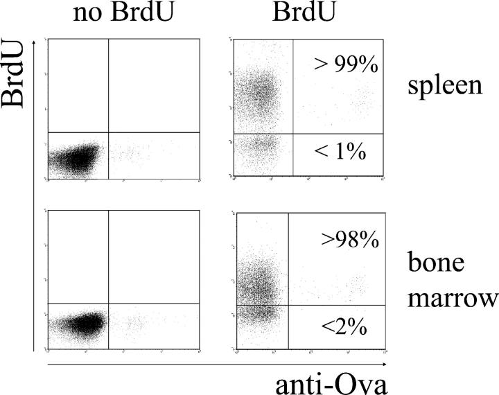
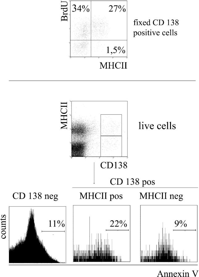
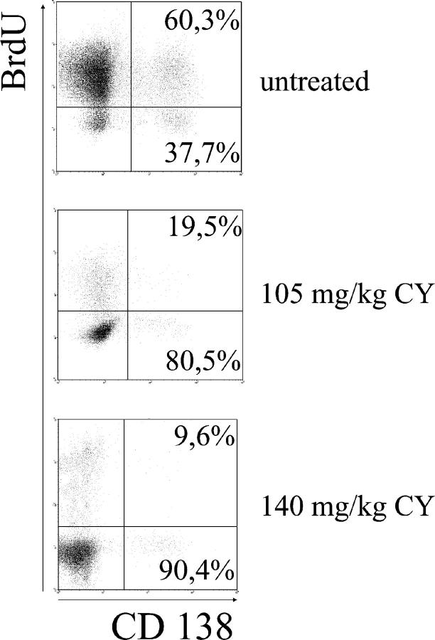
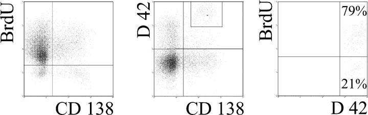
The current view holds that chronic autoimmune diseases are driven by the continuous activation of autoreactive B and T lymphocytes. However, despite the use of potent immunosuppressive drugs designed to interfere with this activation the production of autoantibodies often persists and contributes to progression of the immunopathology. In the present study, we analyzed the life span of (auto)antibody-secreting cells in the spleens of NZB x NZW F1 (NZB/W) mice, a murine model of systemic lupus erythematosus. The number of splenic ASCs increased in mice aged 1-5 mo and became stable thereafter. Less than 60% of the splenic (auto)antibody-secreting cells were short-lived plasmablasts, whereas 40% were nondividing, long-lived plasma cells with a half-life of >6 mo. In NZB/W mice and D42 Ig heavy chain knock-in mice, a fraction of DNA-specific plasma cells were also long-lived. Although antiproliferative immunosuppressive therapy depleted short-lived plasmablasts, long-lived plasma cells survived and continued to produce (auto)antibodies. Thus, long-lived, autoreactive plasma cells are a relevant target for researchers aiming to develop curative therapies for autoimmune diseases.


Figures

Figure 1.
Figure 1.
Kinetics and incorporation of BrdU in the splenic plasma cell compartment of NZB/W mice. (A) Total number of ASCs in spleen, as determined by ELISPOT, at the times indicated. Some groups were fed BrdU continuously (B). Splenic CD138-positive cells consist of dividing and nondividing cells. NZB/W mice with confirmed SLE were continuously fed BrdU for 2 wk. CD138-positive cells were then FACS® sorted and stained for intracellular immunoglobulin (red) and BrdU (green/yellow); original magnification, 160 × (left) and 80 × (right). Plasma cells negative for BrdU had not undergone DNA synthesis or replication during the period of BrdU treatment. Dividing plasmablasts and plasma cells recently derived from proliferating precursors are BrdU positive.
Figure 2.
Figure 2.
The splenic plasma cell compartment consists of continuously dividing cells and a stable population of nondividing cells. NZB/W mice with confirmed SLE were continuously fed BrdU. Splenic plasma cells (CD138-positive) were enumerated by flow cytometry at various sampling times. (A) FACS® analysis. Plasma cells were detected by CD138 staining. Debris was excluded from the analysis by electronic gating. (B) Kinetics of BrdU incorporation in splenic plasma cells based on FACS® analysis. Mean values for five mice are shown. The results are those from one run of two performed. (C) The absolute number of BrdU-negative, nondividing plasma cells was calculated based on their frequency and the absolute number of splenic cells. Each dot resembles an individual mouse.
Figure 3.
Figure 3.
BrdU efficiently labels all newly formed plasma cells. Mice were continuously fed BrdU and immunized and boosted with OVA 3–4 wk later. After fixation, intracellular staining with fluorochrome-labeled OVA was performed to identify plasma cells secreting OVA-specific antibodies. OVA-specific plasma cells in the spleen (top) and BM (bottom) were enumerated 6, 9, and 12 wk after boost immunization. During the whole period between boost immunization and analysis, mice were continuously fed with BrdU. Representative data, as shown, are obtained 6 wk after boost immunization when BrdU incorporation into OVA-specific plasma cells was already complete (n = 5). Similar results were obtained 9 and 12 wk after immunization.
Figure 4.
Figure 4.
Increased binding of early MHCII-positive plasma cells to annexin V. Fixed cells from mice fed with BrdU for 2 wk were stained for CD138, BrdU, and MHCII. Result is shown on cells gated for CD138 expression (top). Frequencies of BrdU-positive, MHCII-negative CD138-positive cells were 32.5 ± 9% (n = 5). Living early plasma cells were distinguished from mature plasma cells by MHCII expression (below) and were stained for annexin V. Dead cells and debris were excluded according to propidium iodide staining and forward scatter profile. Histogram plots were additionally gated on CD138-negative, CD138-positive/MHCII-negative or CD138-positive/MHCII-positive cells.
Figure 5.
Figure 5.
Long-lived plasma cells in NZB/W mice are resistant to conventional immunotherapy. Mice were fed BrdU for 2–4 wk and received the total cyclophosphamide dose indicated (divided over 3 d) 1 wk before sampling. BrdU-positive (short-lived) and -negative (long-lived) plasma cells were identified by FACS®. Representative data for 25 untreated and 12 treated mice are shown.
Figure 6.
Figure 6.
Autoreactive cells can enter the long-lived plasma cell compartment. D42 knock-in mice with an NZB/W genetic background were fed BrdU for 12 wk. The spleens of the mice were then analyzed by FACS® for the presence of BrdU-negative plasma cells (left). D42 transgene-bearing plasma cells were detected by CD138 and intracellular staining with an VH11-specific anti-idiotypic antibody. On the NZB/W background, ∼90% of cells detected with that reagent bind to DNA with high affinity (24) (middle). Gating on D42-positive/CD138-positive plasma cells as shown allowed the identification of BrdU-positive and BrdU-negative plasma cells expressing the D42 transgene (right). Results are representative data from four mice.

Similar articles

Cited by

References

    1. Tan, E.M. 1991. Autoantibodies in pathology and cell biology. Cell. 67:841–842. - PubMed
    1. Ji, H., A.S. Korganow, S. Mangialaio, P. Hoglund, I. Andre, F. Luhder, A. Gonzalez, L. Poirot, C. Benoist, and D. Mathis. 1999. Different modes of pathogenesis in T-cell-dependent autoimmunity: clues from two TCR transgenic systems. Immunol. Rev. 169:139–146. - PubMed
    1. Hahn, B.H. 1998. Antibodies to DNA. N. Engl. J. Med. 338:1359–1368. - PubMed
    1. Lipsky, P.E. 2001. Systemic lupus erythematosus: an autoimmune disease of B cell hyperactivity. Nat. Immunol. 2:764–766. - PubMed
    1. Duke-Cohan, J.S., A. Rubinow, R. Hirt, and D. Naor. 1990. The reaction against autologous lymphoblasts as an indicator of lymphocyte hyperreactivity in rheumatoid arthritis. Clin. Immunol. Immunopathol. 54:298–308. - PubMed

Publication types