. 2004 Jun 7;199(11):1567-75.
doi: 10.1084/jem.20032058. Epub 2004 Jun 1.

Immune responses in healthy and allergic individuals are characterized by a fine balance between allergen-specific T regulatory 1 and T helper 2 cells

Affiliations

Immune responses in healthy and allergic individuals are characterized by a fine balance between allergen-specific T regulatory 1 and T helper 2 cells

Mübeccel Akdis et al. J Exp Med. .

Abstract

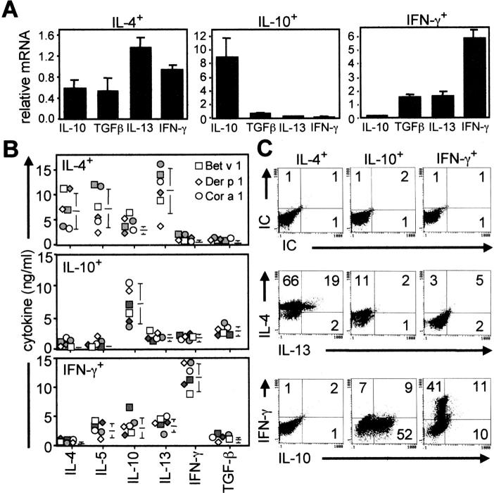
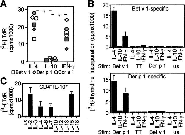
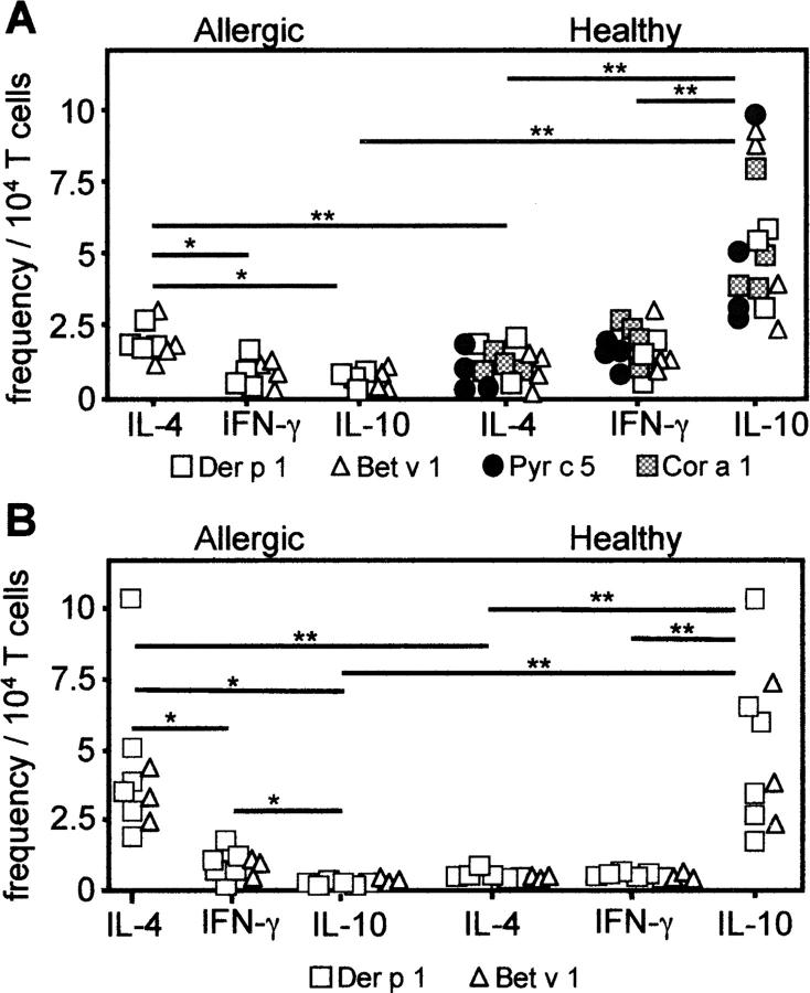
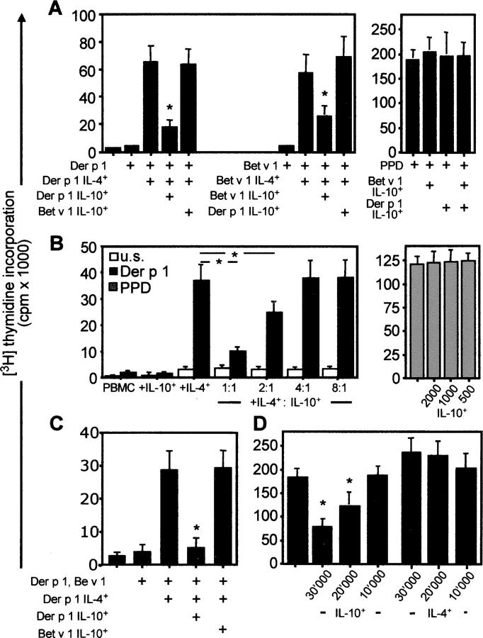
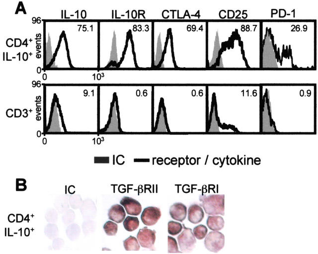
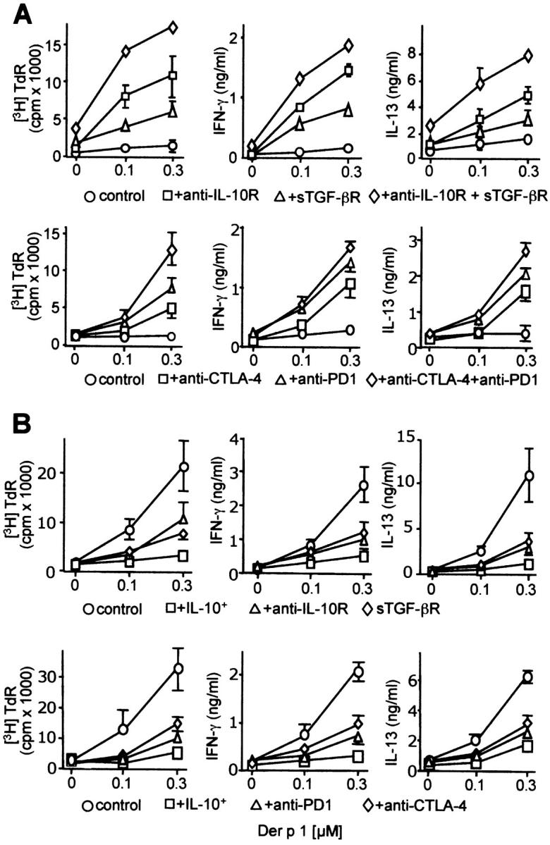
The mechanisms by which immune responses to nonpathogenic environmental antigens lead to either allergy or nonharmful immunity are unknown. Single allergen-specific T cells constitute a very small fraction of the whole CD4+ T cell repertoire and can be isolated from the peripheral blood of humans according to their cytokine profile. Freshly purified interferon-gamma-, interleukin (IL)-4-, and IL-10-producing allergen-specific CD4+ T cells display characteristics of T helper cell (Th)1-, Th2-, and T regulatory (Tr)1-like cells, respectively. Tr1 cells consistently represent the dominant subset specific for common environmental allergens in healthy individuals; in contrast, there is a high frequency of allergen-specific IL-4-secreting T cells in allergic individuals. Tr1 cells use multiple suppressive mechanisms, IL-10 and TGF-beta as secreted cytokines, and cytotoxic T lymphocyte antigen 4 and programmed death 1 as surface molecules. Healthy and allergic individuals exhibit all three allergen-specific subsets in different proportions, indicating that a change in the dominant subset may lead to allergy development or recovery. Accordingly, blocking the suppressor activity of Tr1 cells or increasing Th2 cell frequency enhances allergen-specific Th2 cell activation ex vivo. These results indicate that the balance between allergen-specific Tr1 cells and Th2 cells may be decisive in the development of allergy.


Figures

Figure 1.
Figure 1.
Allergen-specific IL-4-, IFN-γ–, and IL-10–secreting T cells represent Th2-, Th1-, and Tr1-like cells. (A) IL-10, IL-13, IFN-γ, and TGF-β mRNA were quantified by real-time PCR immediately after isolation of Der p 1– or Bet v 1–specific, cytokine-secreting T cells and their relative expression compared with the housekeeping gene EF-1α. The same results were obtained in three independent experiments. (B) Bet v 1–, Der p 1–, and Cor a 1–specific IL-4–, IFN-γ–, and IL-10–secreting T cells (one allergic and one healthy donor each; closed symbols, allergic donors; open symbols, healthy donors) were in vitro expanded for 2 wk, and their cytokine profile was determined in supernatants by ELISA 72 h after anti-CD2, anti-CD3, and anti-CD28 mAb stimulation. (C) Intracytoplasmic cytokine profile of in vitro–expanded allergen-specific T cells (the same results were obtained in 10 additional experiments). Percentage of positive cells is shown in each quadrant.
Figure 2.
Figure 2.
Specificity and growth requirements of purified allergen-specific T cells. (A) Bet v 1–, Der p 1–, and Cor a 1–specific IL-4–, IFN-γ–, and IL-10–secreting T cells (one allergic and one healthy donor each; closed symbols, allergic donors; open symbols, healthy donors) were purified and in vitro expanded, and 5 × 10⁵ cells were cocultured with 5 × 10⁵ autologous irradiated PBMCs as an APC source in the presence of the same antigen. [3H]Thymidine incorporation (TdR) was determined after 5 d. *, P < 0.001. (B) Purified Bet v 1–specific and Der p 1–specific T cells did not show any proliferative response to different control antigens (two representative of six experiments are shown; mean ± SD of triplicate cultures). TT, tetanus toxoid; us, unstimulated. (C) Growth factor requirements of antigen-specific IL-10–secreting T cells. After purification, Bet v 1– or Der p 1–specific IL-10–secreting T cells were first expanded for 10 d in the presence of IL-2, washed, and cultured (5 × 10⁴ cells) with 1-nM doses of different cytokines. [3H]Thymidine incorporation was determined after 3 d (mean ± SD of three independent experiments are shown).
Figure 3.
Figure 3.
Increased frequency of allergen-specific Tr1 cells in healthy individuals and Th2 cells in allergic individuals. (A) Frequency of Der p 1– and Bet v 1–specific, cytokine-secreting CD4+ T cells from eight allergic individuals and Cor a 1–, Bet v 1-, Der p 1–, and Pyr c 5–specific, cytokine-secreting CD4+ T cells from 15 healthy individuals out of pollen season. (B) Frequency of Der p 1–specific T cells in six allergic and six healthy, and Bet v 1–specific T cells in three allergic and three healthy individuals by ELISPOT assay. *, P < 0.01; **, P < 0.001.
Figure 4.
Figure 4.
Antigen-specific suppression by Tr1 cells. (A) Der p 1–specific and Bet v 1–specific IL-4–secreting and IL-10–secreting T cells were purified from healthy individuals. Their frequency was calculated in CD4+ T cells, and 2 × 10⁵ PBMCs were immediately reconstituted by increasing their frequency by 10 times (IL-4–secreting T cells, 0.02–0.2%; IL-10–secreting T cells, 0.05–0.5%). Cells were stimulated with the respective antigens and PPD. (B) Der p 1–specific IL-4–secreting and IL-10–secreting T cells were purified and in vitro expanded by IL-2/IL-4 and IL-2/IL-15, respectively. 2 × 10⁵ PBMCs were enriched with 1,000 Der p 1–specific IL-4– or IL-10–secreting T cells or their combinations in IL-4–secreting/IL-10–secreting T cell ratios 1,000/1,000 (1:1), 1,000/500 (2:1), 1,000:250 (4:1), and 1,000:125 (8:1). Cells were stimulated with 0.3 μM Der p 1 or 1 μg/ml PPD. Der p 1–specific, IL-10–secreting T cells added to PPD-stimulated PBMC cultures at indicated numbers did not show any suppression. (C) The same experimental design as in A was used, and cells were stimulated with both Der p 1 and Bet v 1 (0.3 μM each). (A–C) [3H]Thymidine incorporation was determined after 5 d. The same results were obtained in three other experiments. *, P < 0.001. (D) 105 PBMCs were stimulated with anti-CD3 in the presence of different amounts of IL-10–secreting and IL-4-secreting T cells. [3H]Thymidine incorporation was determined after 3 d. Data represent two different experiments. *, P < 0.01.
Figure 5.
Figure 5.
Expression of suppressor molecules on IL-10–secreting T cells. (A) Immediately after purification, antigen-specific IL-10–secreting T cells of healthy individuals were analyzed for intracytoplasmic IL-10, surface IL-10R, CTLA-4, CD25, and PD-1 by flow cytometry. Data are compared with CD3+ T cells of the same donor stimulated with the same antigen. (B) TGF-β RI and RII expression by immunohistology. (A and B) Same results were obtained in three independent experiments.
Figure 6.
Figure 6.
Multiple suppressive mechanisms play a role in peripheral allergen tolerance. (A) Endogenous IL-10, TGF-β, or both as well as CTLA-4, PD-1, or both were neutralized in Der p 1–stimulated PBMCs of healthy individuals. [3H]Thymidine incorporation (TdR), IFN-γ, and IL-13 were determined at day 5. (B) Der p 1–specific proliferation of PBMCs from house dust mite allergic patients was suppressed by 10 times increased frequency of Der p 1–specific, IL-10–secreting T cells (IL-10+). The activity of IL-10, TGF-β, CTLA-4, and PD-1 were neutralized. [3H]Thymidine incorporation (TdR), IFN-γ, and IL-13 were determined at day 5. (control) Isotype control antibody. (A and B) Same results were obtained in four independent experiments (two Bet v1 and two Der p 1) all performed with freshly purified cells without in vitro expansion. Bet v 1 stimulation in birch pollen–allergic individuals gave similar results. All of the neutralization experiments were significantly different from the control and IL-10–secreting T cell–suppressed condition at 0.3 μM allergen doses. P < 0.001.

Similar articles

Cited by

References

    1. Teale, J.M., and N.R. Klinman. 1980. Tolerance as an active process. Nature. 288:385–387. - PubMed
    1. Parijs, L.V., and A.K. Abbas. 1998. Homeostasis and self-tolerance in the immune system: turning lymphocytes off. Science. 280:243–247. - PubMed
    1. Mosmann, T.R., H. Cherwinski, M.W. Bond, M.A. Giedlin, and R.L. Coffman. 1986. Two types of murine helper T cell clones. 1. Definition according to profiles of lymphokine activities and secreted proteins. J. Immunol. 136:2348–2357. - PubMed
    1. Romagnani, S. 1994. Lymphokine production by human T cells in disease states. Annu. Rev. Immunol. 12:227–257. - PubMed
    1. Gershon, R.K., and K. Kondo. 1970. Cell interactions in the induction of tolerance: the role of thymic lymphocytes. Immunology. 18:723–737. - PMC - PubMed

Publication types