

Classical Article
doi: 10.1085/jgp.200409091.
The early history of the biochemistry of muscle contraction
- PMID: 15173217
- PMCID: PMC2234565
- DOI: 10.1085/jgp.200409091
Item in Clipboard
Classical Article
The early history of the biochemistry of muscle contraction
J Gen Physiol.
2004 Jun.
No abstract available
Figures
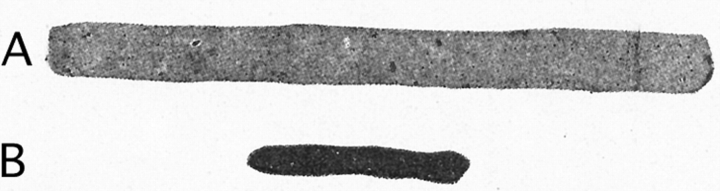
Contraction of actomyosin threads (“myosin B”) on addition of ATP. Shown are the same thread A before and B after addition of boiled muscle juice (a source of ATP) (Szent-Györgyi, 1942a).
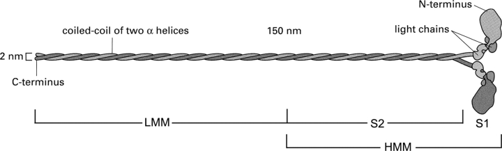
The myosin molecule (adapted from Alberts et al., 2002).
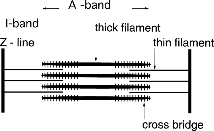
Cross-striated muscle is organized in sarcomeres that extend from one Z-line to the next. The distance between Z-lines is 2–3 μm. The thin filaments contain actin and the thick filaments contain myosin. The thick filaments have bipolar symmetry with a central bare zone in which there are no cross-bridges. The actin fiber symmetry reverses in the Z-line. The area not penetrated by the thin filaments is variable, and is known as the H-zone.
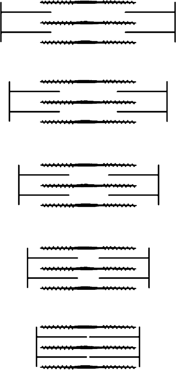
2-μm long myosin containing thick filaments with cross-bridges and 1-μm long actin containing thin filaments are shown. As the sarcomere shortens, the myosin cross-bridges react with actin and propel the thin filaments toward the center of the sarcomere. Both filament types remain at constant lengths during contraction. The sliding of the filaments explains the constancy of the A-band and the changes of the I-band and the H-zone.
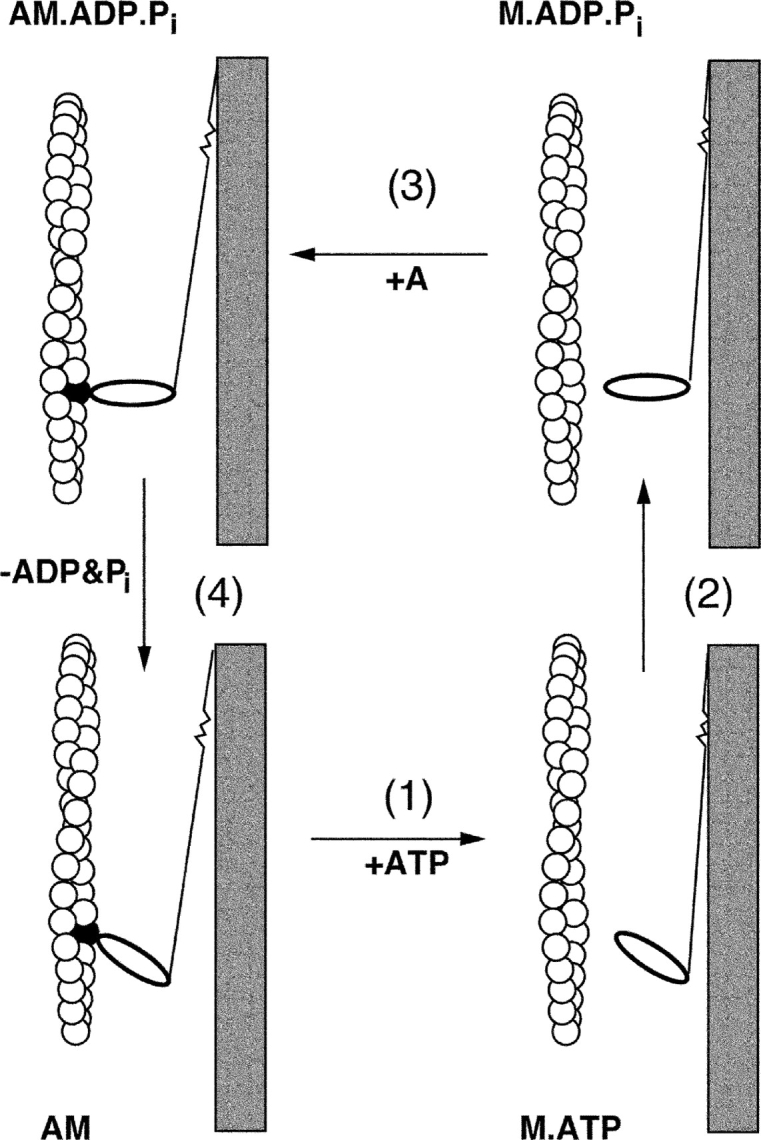
The cross-bridge cycle. Note that ATP hydrolysis takes place in the detached state. In the actin-bound state contraction is associated with the dissociation of the hydrolysis products; recovery of the resting state structure follows dissociation of myosin from actin by ATP (see Taylor, 2001).
Similar articles
-
Conformational switching in muscle.Adv Exp Med Biol. 2004;547:61-80. doi: 10.1007/978-1-4419-8861-4_6. Adv Exp Med Biol. 2004. PMID: 15230093 Review.
-
Functional classification of skeletal muscle networks. I. Normal physiology.J Appl Physiol (1985). 2012 Dec 15;113(12):1884-901. doi: 10.1152/japplphysiol.01514.2011. Epub 2012 Oct 18. J Appl Physiol (1985). 2012. PMID: 23085959 Free PMC article.
-
Molecules in motion: Michael Sheetz, James Spudich, and Ronald Vale receive the 2012 Albert Lasker Basic Medical Research Award.J Clin Invest. 2012 Oct;122(10):3374-7. doi: 10.1172/jci66361. J Clin Invest. 2012. PMID: 23193575 Free PMC article. No abstract available.
-
Memories of Annemarie Weber.Anat Rec (Hoboken). 2014 Sep;297(9):1543-7. doi: 10.1002/ar.22963. Anat Rec (Hoboken). 2014. PMID: 25125168 Free PMC article. No abstract available.
-
Lifting the nebula: novel insights into skeletal muscle contractility.Physiology (Bethesda). 2010 Oct;25(5):304-10. doi: 10.1152/physiol.00016.2010. Physiology (Bethesda). 2010. PMID: 20940435 Review.
Cited by
-
Protein nanomechanics in biological context.Biophys Rev. 2021 Aug 7;13(4):435-454. doi: 10.1007/s12551-021-00822-9. eCollection 2021 Aug. Biophys Rev. 2021. PMID: 34466164 Free PMC article. Review.
-
Thick Filament Protein Network, Functions, and Disease Association.Compr Physiol. 2018 Mar 13;8(2):631-709. doi: 10.1002/cphy.c170023. Compr Physiol. 2018. PMID: 29687901 Free PMC article. Review.
-
Order-Disorder Transitions in the Cardiac Troponin Complex.J Mol Biol. 2016 Jul 31;428(15):2965-77. doi: 10.1016/j.jmb.2016.06.022. Epub 2016 Jul 6. J Mol Biol. 2016. PMID: 27395017 Free PMC article. Review.
-
Use of Amino Acids as Supplements for Matching Nutrition, Training, and Rehabilitation-Focusing on Some Questions.Nutrients. 2025 Aug 18;17(16):2667. doi: 10.3390/nu17162667. Nutrients. 2025. PMID: 40871694 Free PMC article. Review.
-
Reconstitution of contractile actomyosin rings in vesicles.Nat Commun. 2021 Apr 15;12(1):2254. doi: 10.1038/s41467-021-22422-7. Nat Commun. 2021. PMID: 33859190 Free PMC article.
References
-
- Alberts, B., A. Johnson, J. Lewis, M. Raff, K. Roberts and P. Walter. 2002. Molecular Biology of the Cell. Fourth edition. Garland Publishing. 950 pp.
-
- Asakura, S. 1961. F-actin adenosine triphosphatase activated under sonic vibration. Biochim. Biophys. Acta. 52:65–75. - PubMed
-
- Asakura, S., M. Taniguchi, and F. Oosawa. 1963. Mechano-chemical behaviour of F-actin. J. Mol. Biol. 7:55–69.
-
- Astbury, W.T. 1947. Croonian lecture. On the structure of biological fibers and the problem of muscle. Proc. R. Soc. Lond. B. Biol. Sci. 134:303–328. - PubMed
