. 2004 Jun 8;101(23):8664-9.
doi: 10.1073/pnas.0402354101. Epub 2004 Jun 1.

Bardet-Biedl syndrome type 4 (BBS4)-null mice implicate Bbs4 in flagella formation but not global cilia assembly

Affiliations

Bardet-Biedl syndrome type 4 (BBS4)-null mice implicate Bbs4 in flagella formation but not global cilia assembly

Kirk Mykytyn et al. Proc Natl Acad Sci U S A. .

Abstract

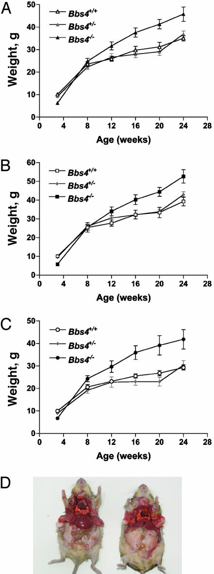
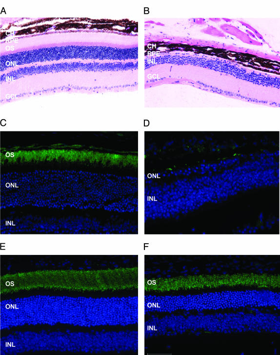
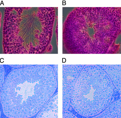
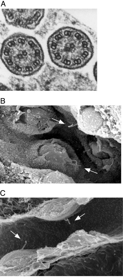
The functions of the proteins encoded by the Bardet-Biedl syndrome (BBS) genes are unknown. Mutations in these genes lead to the pleiotropic human disorder BBS, which is characterized by obesity, retinopathy, polydactyly, renal and cardiac malformations, learning disabilities, and hypogenitalism. Secondary features include diabetes mellitus and hypertension. Recently, it has been suggested that the BBS phenotypes are the result of a lack of cilia formation or function. In this study, we show that mice lacking the Bbs4 protein have major components of the human phenotype, including obesity and retinal degeneration. We show that Bbs4-null mice develop both motile and primary cilia, demonstrating that Bbs4 is not required for global cilia formation. Interestingly, male Bbs4-null mice do not form spermatozoa flagella, and BBS4 retinopathy involves apoptotic death of photoreceptors, the primary ciliated cells of the retina. These mutation data demonstrate a connection between the function of a BBS protein and cilia. To further evaluate an association between cilia and BBS, we performed homology comparisons of BBS proteins in model organisms and find that BBS proteins are specifically conserved in ciliated organisms.


Figures

Fig. 1.
Fig. 1.
Targeting of the Bbs4 gene. (A) Diagram of the Bbs4 targeting vector and the resulting recombination product. Upon homologous recombination, exons 6–11 are replaced with a neomycin cassette. (B)(Upper) Northern blot analysis of Bbs4 expression in kidney total cellular RNA from wild-type (+/+), heterozygous (+/–), and homozygous (–/–) animals. The probe is a 960-bp Bbs4 partial 3′ cDNA. (Lower) The same blot probed with β-actin as a loading control.
Fig. 2.
Fig. 2.
Comparison of weight and adipose tissue deposition in Bbs4 mice. (A) The weights of all wild-type (Bbs4+/+; n = 40), heterozygous (Bbs4+/–; n = 40), and homozygous (Bbs4–/–; n = 36) mice at 3, 8, 12, 16, 20, and 24 weeks of age. Values are expressed as mean ± SEM. (B) The weights of male wild-type (Bbs4+/+; n = 20), heterozygous (Bbs4+/–; n = 20), and homozygous (Bbs4–/–; n = 20) mice. Values are expressed as mean ± SEM. (C) The weights of female wild-type (Bbs4+/+; n = 20), heterozygous (Bbs4+/–; n = 20), and homozygous (Bbs4–/–; n = 16) mice. Values are expressed as mean ± SEM. (D) Examination of adipose tissue deposition demonstrates an increase in the amount of central adipose tissue in a Bbs4–/– mouse (Right), as compared to a Bbs4+/+ mouse (Left). The mice are ≈10-month-old males.
Fig. 3.
Fig. 3.
Examination of mouse retina. (A and B) Hematoxylin and eosin staining of retinal sections from a Bbs4+/+ (A) and Bbs4–/– (B) mouse. The entire photoreceptor layer is absent in the Bbs4–/– section. CH, choroids; RPE, retinal pigment epithelium; OS, outer segment; ONL, outer nuclear layer; INL, inner nuclear layer; GCL, ganglion cell layer. (C and D) Labeling of rods with anti-Opsin (green) from a Bbs4+/+ (C) and Bbs4–/– (D) mouse. The lack of labeling in the Bbs4–/– retinal section confirms the absence of the photoreceptors. (E and F) Labeling of rods with anti-Opsin (green) from a 6-week-old Bbs4+/+ (E) and Bbs4–/– (F) mouse, demonstrating the presence of photoreceptors. The outer nuclear and photoreceptor layers in the Bbs4–/– section are approximately one-half the thickness of the Bbs4+/+ section, indicating significant photoreceptor cell degeneration.
Fig. 4.
Fig. 4.
Histological examination of mouse seminiferous tubules. (A) Hematoxylin and eosin staining of a testes section from a Bbs4+/+ mouse. Flagella are apparent in the lumen of the tubule. (B) Hematoxylin and eosin staining of a testes section from a Bbs4–/– mouse showing the absence of flagella in the lumen. (C) Toluidine blue staining of a 1-μm testes section from a Bbs4+/+ mouse showing spermatozoa with normal morphology as evidenced by the presence of flagella and condensed sperm nuclei. (D) Toluidine blue staining of a 1-μm testes section from a Bbs4–/– mouse showing the presence of condensed sperm nuclei in the absence flagella.
Fig. 5.
Fig. 5.
Electron microscopy analysis of motile and primary cilia. (A) Transmission electron microscopic image of a cross-section of cilia in the trachea of a Bbs4–/– mouse. The nine outer and two inner microtubules are clearly defined, indicating that the internal structure of motile cilia appears normal. (B and C) Scanning electron microscopic image of renal tubule cells from a Bbs4+/+ (B) and a Bbs4–/– (C) mouse. Normal appearing primary cilia (arrows) are evident in both.

References

    1. Mykytyn, K., Nishimura, D. Y., Searby, C. C., Shastri, M., Yen, H. J., Beck, J. S., Braun, T., Streb, L. M., Cornier, A. S., Cox, G. F., et al. (2002) Nat. Genet. 31, 435–438. - PubMed
    1. Nishimura, D. Y., Searby, C. C., Carmi, R., Elbedour, K., Van Maldergem, L., Fulton, A. B., Lam, B. L., Powell, B. R., Swiderski, R. E., Bugge, K. E., et al. (2001) Hum. Mol. Genet. 10, 865–874. - PubMed
    1. Mykytyn, K., Braun, T., Carmi, R., Haider, N. B., Searby, C. C., Shastri, M., Beck, G., Wright, A. F., Iannaccone, A., Elbedour, K., et al. (2001) Nat. Genet. 28, 188–191. - PubMed
    1. Slavotinek, A. M., Stone, E. M., Mykytyn, K., Heckenlively, J. R., Green, J. S., Heon, E., Musarella, M. A., Parfrey, P. S., Sheffield, V. C. & Biesecker, L. G. (2000) Nat. Genet. 26, 15–16. - PubMed
    1. Katsanis, N., Beales, P. L., Woods, M. O., Lewis, R. A., Green, J. S., Parfrey, P. S., Ansley, S. J., Davidson, W. S. & Lupski, J. R. (2000) Nat. Genet. 26, 67–70. - PubMed

Publication types

MeSH terms