. 2004 Jun;113(11):1589-95.
doi: 10.1172/JCI21334.

Concentration-dependent regulation of thyrotropin receptor function by thyroid-stimulating antibody

Affiliations

Concentration-dependent regulation of thyrotropin receptor function by thyroid-stimulating antibody

Takao Ando et al. J Clin Invest. 2004 Jun.

Abstract

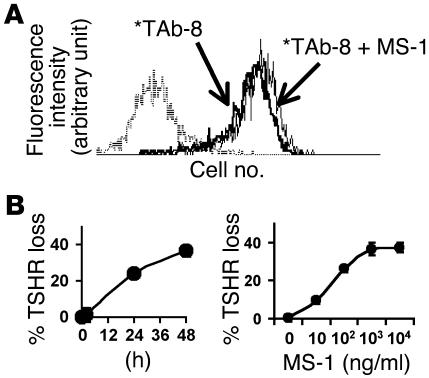
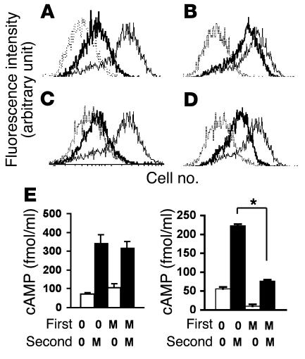
Thyrotropin receptor (TSHR) Ab's of the stimulating variety are the cause of hyperthyroid Graves disease. MS-1 is a hamster mAb with TSHR-stimulating activity. To examine the in vivo biological activity of MS-1, mice were treated with purified MS-1 intraperitoneally and the thyroid response evaluated. MS-1 induced a dose-dependent increase in serum thyroxine (T4), with a maximum effect after 10 proportional, variant g of MS-1 was administered. MS-1-secreting hybridoma cells were then transferred into the peritoneum of nude mice to study chronic thyroid stimulation. Serum MS-1 levels detected after 2 weeks were approximately 10-50 proportional, variant g/ml, and the serum TSH was suppressed in all animals. Serum triiodothyronine levels were elevated, but only in animals with low serum MS-1 concentrations. In addition, there was a negative correlation between serum T4 and the serum MS-1 concentrations. These in vivo studies suggested a partial TSHR inactivation induced by excessive stimulation by MS-1. We confirmed this inactivation by demonstrating MS-1 modulation of TSHR function in vitro as evidenced by downregulation and desensitization of the TSHR at concentrations of MS-1 achieved in the in vivo studies. Thus, inactivation of the TSHR by stimulating TSHR autoantibodies (TSHR-Ab's) in Graves disease patients may provide a functional explanation for the poor correlation between thyroid function and serum TSHR-Ab concentrations.


Figures

Figure 1
Figure 1
Mouse TSHR binding and stimulation by MS-1 in vitro. (A) Binding of MS-1 on CHO-mTSHR cells (solid line) and nonspecific binding to control CHO cells (broken line), expressed as mean fluorescence intensity (MFI). (B) CHO-mTSHR cells were stimulated for 2 hours with indicated concentrations of MS-1. Significant stimulation (more than twofold cAMP production compared with basal) was obtained with less than 10 ng/ml MS-1.
Figure 2
Figure 2
Acute thyroid stimulation in vivo by MS-1. (A) CBA/J mice were injected with the indicated doses of MS-1 or control (Ctl) Ab, and T4 levels were measured every 24 hours. A broken line indicates the upper level for control serum T4. *P < 0.01 in T4 levels between 48 hours and 72 hours with a 10-∝g injection. (B) Area under the curve (AUC) of the T4 released in response to indicated doses of MS-1 stimulation. #P < 0.002 and **P < 0.0001 in AUC compared with the control treatment. (C) Example of thyroid histology at 96 hours in control (upper) and MS-1 (10 ∝g) treated animals (lower). Note thyroid epithelial cell hypertrophy. Magnification, ∞400.
Figure 3
Figure 3
Chronic thyroid stimulation in vivo by MS-1. (A and B) Correlation between serum MS-1 levels and (A) T3 and (B) T4 concentrations. The gray areas indicate normal ranges (mean ± 2SE). Data for animals with thyroid hypertrophy and atrophy were expressed as filled circles and filled triangles, respectively. (C_F) Thyroid glands from nude mice, showing (C) thyroid epithelial hypertrophy with vascular engorgement (D) thyroid epithelial hypertrophy with colloid depletion (E) thyroid atrophy, and (F) a normal thyroid. Magnification, ∞200. (G) Treated mouse serum bound to CHO-mTSHR (thick line) and not to control CHO cells (thin line). Anti-hamster IgG and anti-mouse IgG (inset) were used to detect IgG bound to the cells. The horizontal axis indicates fluorescence intensity and the vertical axis the cell number. (H) Representative serum from an MS-1 hybridoma_treated nude mouse containing approximately 50 ∝g/ml of MS-1 was serially diluted and used for stimulation of CHO-mTSHR cells.
Figure 4
Figure 4
TSHR stimulation study in vitro by MS-1. (A) TSHR activation profile in vitro at different time points. CHO-hTSHR cells were stimulated with indicated doses of MS-1 for indicated periods. *P < 0.05. (B) TSHR responsiveness with 48 hours of stimulation by MS-1. A ratio for cAMP production with indicated doses of MS-1 (2 hours vs. 48 hours; inset) was expressed as percentage responsiveness. * P < 0.05; **P < 0.02 (indicated dose vs. 0, 10, and 33 ng/ml of MS-1). (C) MS-1 binding when loss of TSHR responsiveness was observed. CHO-hTSHR cells were stimulated with the indicated doses of MS-1 for 48 hours, and surface MS-1 binding was studied. MS-1 binding (mean ± SD) in five separate experiments on CHO-hTSHR cells without prior MS-1 stimulation is shown by the arrows.
Figure 5
Figure 5
TSHR downregulation by MS-1. (A) Binding of labeled TAb-8 (*TAb-8) to CHO-hTSHR in the presence (thin line) or absence (thick line) of MS-1. The broken line indicates background staining. (B) TSHR downregulation. Time dependency in response to 1 ∝g/ml of MS-1 (left) and dose dependency with 48 hours of MS-1 stimulation (right). Loss of cell surface TSHRs was detected by labeled TAb-8, expressed as percentage TSHR loss after 48 hours. The use of TAb-8 avoided the problem of receptor occupancy by MS-1.
Figure 6
Figure 6
TSHR desensitization by MS-1. (A_D) Optimization of acid wash. MS-1 binding to CHO-hTSHR cells (thin solid lines), bound MS-1 (thick line) after acid wash for 30 seconds (A) or 4 minutes (C), and rebinding of MS-1 (thick lines) after acid wash for 30 seconds (B) or 4 minutes (D) are shown. Broken lines indicate background staining. (E) TSHR desensitization. CHO-hTSHR cells were stimulated with 1 ∝g/ml of MS-1 or medium (indicated as M and 0, respectively) for 4 hours (left) and 48 hours (right) (first stimulation), followed by 2 hours incubation with or without the same dose of MS-1 with 2 mM isobutylmethyl-xanthine (second stimulation). Between these two stimuli, cells were washed with cold acid buffer. Forskolin (50 ∝M) stimulation generated approximately 3,000 fmol of cAMP. *P < 0.001.

References

    1. Davies T, Marians R, Latif R. The TSH receptor reveals itself. J. Clin. Invest. 2002;110:161–164. doi:10.1172/JCI200216234. - PMC - PubMed
    1. Rees Smith B, McLachlan SM, Furmaniak J. Autoantibodies to the thyrotropin receptor. Endocr. Rev. 1988;9:106–121. - PubMed
    1. Rapoport B, Chazenbalk GD, Jaume JC, McLachlan SM. The thyrotropin (TSH) receptor: interaction with TSH and autoantibodies. Endocr. Rev. 1998;19:673–716. - PubMed
    1. Adams DD, Purves HD. Abnormal responses in the assay of thyrotropins. Proc. Univ. Otago Med. Sch. 1956;34:11–12.
    1. Kriss J, Pleshakov V, Chien JR. Isolation and identification of the long acting thyroid stimulator and its relation to hyperthyroidism and circumscribed pretibial myxedema. J. Clin. Endocrinol. Metab. 1964;24:1005–1028. - PubMed

Publication types

Substances