. 2004 Jun;113(11):1641-50.
doi: 10.1172/JCI18704.

Receptor for advanced glycation end products (RAGE) regulates sepsis but not the adaptive immune response

Affiliations

Receptor for advanced glycation end products (RAGE) regulates sepsis but not the adaptive immune response

Birgit Liliensiek et al. J Clin Invest. 2004 Jun.

Abstract

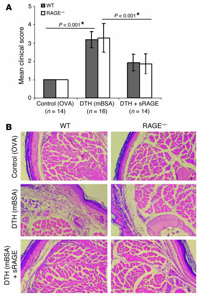
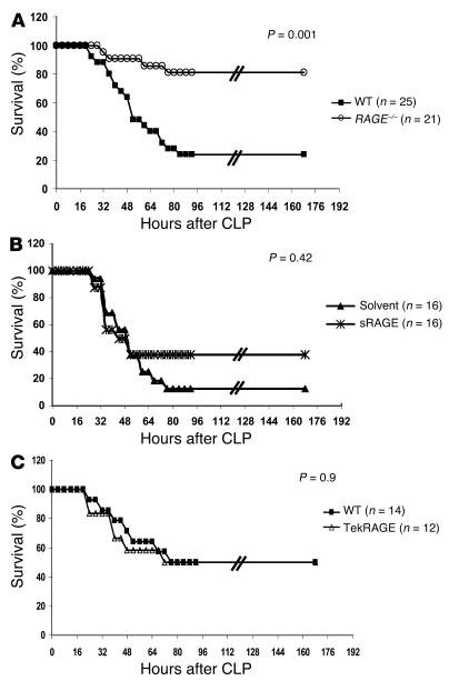
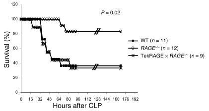
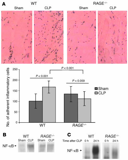
While the initiation of the adaptive and innate immune response is well understood, less is known about cellular mechanisms propagating inflammation. The receptor for advanced glycation end products (RAGE), a transmembrane receptor of the immunoglobulin superfamily, leads to perpetuated cell activation. Using novel animal models with defective or tissue-specific RAGE expression, we show that in these animal models RAGE does not play a role in the adaptive immune response. However, deletion of RAGE provides protection from the lethal effects of septic shock caused by cecal ligation and puncture. Such protection is reversed by reconstitution of RAGE in endothelial and hematopoietic cells. These results indicate that the innate immune response is controlled by pattern-recognition receptors not only at the initiating steps but also at the phase of perpetuation.


Figures

Figure 1
Figure 1
Generation and characterization of RAGE–/– mouse line. (A) RT-PCR of lung tissue from RAGE–/– and WT mice analyzed for RAGE mRNA expression. WT indicates RAGE+/+ littermates. HPRT was used to demonstrate equal loading. (B) Western blot analysis of RAGE–/– and WT lung tissue analyzed for the presence of RAGE protein (43 kDa). Tubulin demonstrates equal loading. (C) Immunohistology for RAGE protein in WT and RAGE–/– lung tissue. Brown staining indicates RAGE expression; magnification, ∞400.
Figure 2
Figure 2
Generation and characterization of the Tie2RAGE mouse line. (A) Tie2RAGE construct. Genomic RAGE DNA was cloned between the Tie2 promoter and enhancer. Gray boxes indicate RAGE exons; Roman numerals indicate RAGE exon numbers. E, EcoRI restriction enzyme site. (B) Southern blot analysis of DNA obtained from tail biopsies from WT and Tie2RAGE mice; EcoRI digestion releases a 2.2-kb DNA fragment from the Tie2RAGE transgene. (C) RT-PCR of different tissues derived from Tie2RAGE (Tie2) and WT mice analyzed for RAGE mRNA expression. WT indicates transgene-negative littermates. HPRT was used to demonstrate equal loading. (D) Immunohistology for RAGE protein in Tie2RAGE and WT kidney tissue. Brown staining indicates RAGE expression; magnification, ∞400. RAGE expression in kidneys of Tie2RAGE mice is evident in the endothelium of large and small vessels, while no RAGE protein could be detected in kidneys of healthy WT mice.
Figure 3
Figure 3
NF-κB inducibility in peripheral blood mononuclear cells of the various mouse strains. Six-month-old WT, RAGE–/–, Tie2RAGE, or Tie2RAGE ∞ RAGE–/– mice received unmodified control hemoglobin (Contr.-Hg) or CML-modified hemoglobin (CML-Hg) (1,000 ∝g/mouse at time point 0, intraperitoneally). Six days later, mice were sacrificed and NF-κB–binding activity was determined in total blood. Recombinant NF-κB (Rec. NF-κB) produced in erythrocyte lysates served as control for NF-κB binding. To confirm NF-κB binding, binding activity at 6 days was competed with a 160-fold molar excess of unlabeled consensus NF-κB oligonucleaotides (cons.). Arrowheads indicate NF-κB complexes consisting of NF-κB homodimers or NF-κB (p50/p65) heterodimers, respectively.
Figure 4
Figure 4
RAGE–/– and Tie2RAGE mice develop a normal immune response in EAE. Age- and sex-matched mice were sensitized against oligodendrocyte glycoprotein (MOG35–55) and were monitored for the onset of clinical signs on a daily basis (see Methods). (A) EAE response in WT and RAGE–/– mice. WT indicates C57BL/6 ∞ 129/Sv mice. The mean clinical score represents a summary of two independent experiments each for WT and for RAGE–/–. WT, filled squares; RAGE–/–, open circles. (B) EAE response in WT and Tie2RAGE mice. WT mice used were transgene-negative littermates. The mean clinical score represents a summary of two independent experiments each for WT and for Tie2RAGE. WT, filled squares; Tie2RAGE, open triangles. The standard error of the mean is given (± SEM), and P < 0.05 was considered to be statistically significant.
Figure 5
Figure 5
RAGE–/– mice display normal inflammation in a model of DTH, and application of sRAGE blocks inflammation in DTH in WT and RAGE–/– mice. Age- and sex-matched WT and RAGE–/– mice were sensitized with mBSA. WT indicates C57BL/6 ∞ 129/Sv mice. Control groups were challenged with ovalbumin (OVA), and DTH groups, with mBSA. Mouse groups receiving sRAGE were pretreated by intraperitoneal injection of solvent or sRAGE 24 and 12 hours prior to and 6 and 12 hours after local challenge with mBSA. The DTH experiment was repeated three times and the DTH with sRAGE treatment was repeated two times with similar results (A). The mean clinical inflammation score represents a summary of two independent experiments. Twenty-four hours after footpad injection, mice were subjected to clinical scoring (see Methods). Standard error is given (± SEM), and P < 0.05 was considered to be statistically significant (*). WT, gray bars; RAGE–/–, white bars. (B) Representative pictures of hematoxylin and eosin–stained footpad sections of experimental groups; magnification, ∞400.
Figure 6
Figure 6
RAGE–/– mice and WT mice treated with sRAGE are protected against septic shock induced by CLP. (A) Age- and sex-matched WT and RAGE–/– mice were subjected to CLP or sham operation and were monitored for survival. WT indicates C57BL/6 ∞ 129/Sv mice. A summary of four repeated experiments is shown. One to two sham-operated mice were included in each single experiment (data not shown) and did not show overt signs of disease. WT, filled squares; RAGE–/–, open circles. (B) Therapeutic effect of sRAGE treatment. CLP was performed in age- and sex-matched C57BL/6 mice. Mice received repeated intraperitoneal injections of sRAGE or solvent (LPS-free 0.9% NaCl) immediately after CLP (150 ∝g sRAGE/mouse) and 6, 12, 24, and 36 hours thereafter (70 ∝g sRAGE/mouse). Two repeated experiments are summarized. WT treated with solvent, filled triangles; WT treated with sRAGE, asterisks. (C) Age- and sex-matched WT and Tie2RAGE mice were subjected to CLP and were monitored for survival. WT indicates transgene-negative littermates. A summary of two repeated experiments is shown. One to two sham-operated mice were included in each single experiment (data not shown) and did not show overt signs of disease. WT, filled squares; Tie2RAGE, open triangles.
Figure 7
Figure 7
Rescue of RAGE expression in hematopoietic cells and the endothelium abolishes protection from septic shock independently of phagocytosis after CLP. Age- and sex-matched WT, RAGE–/–, and Tie2RAGE ∞ RAGE–/– mice were subjected to CLP and were monitored for survival. Additionally, one to two sham-operated mice were included in each single experiment (data not shown). WT indicates C57BL/6 ∞ 129/Sv mice. A summary of two repeated experiments is shown. WT, filled squares; RAGE–/–, open circles; Tie2RAGE ∞ RAGE–/–, asterisks.
Figure 8
Figure 8
Inflammatory cell adhesion on the peritoneum and NF-κB activation after CLP is reduced in RAGE–/– mice. Age- and sex-matched WT and RAGE–/– mice were sham-operated (Sham) or were subjected to CLP. WT indicates C57BL/6 ∞ 129/Sv mice. Twenty-four hours after sham operation or CLP, peritoneum was harvested for histology or isolation of nuclear proteins for EMSA. Experiments were repeated three times with similar results. (A) Representative pictures of hematoxylin and eosin–stained peritoneum sections from experimental groups showing adherent inflammatory cells; magnification, ∞400. Cells in visual fields of serial sections of peritoneum were counted using a microscope, and the results were summarized and analyzed statistically. The standard error is given (± SEM), and P < 0.05 was considered to be statistically significant (*). WT, dark gray bars; RAGE–/–, light gray bars. (B) Representative EMSA for NF-κB–binding activity in peritoneum derived from WT and RAGE–/– mice 24 hours after sham operation or CLP. (C) Representative EMSA for NF-κB–binding activity in lungs derived from WT and RAGE–/– mice before (0 h) and 24 hours after (24 h) CLP.

References

    1. Zhang G, Ghosh S. Toll-like receptor-mediated NF-κB activation: a phylogenetically conserved paradigm in innate immunity. J. Clin. Invest. 2001;107:13–19. - PMC - PubMed
    1. Medzhitov R, Preston Hurlburt P, Janeway CA., Jr A human homologue of the Drosophila toll protein signals activation of adaptive immunity. Nature. 1997;388:394–397. - PubMed
    1. Modlin RL, Brightbill HD, Godowski PJ. The toll of innate immunity on microbial pathogens. N. Engl. J. Med. 1999;340:1834–1835. - PubMed
    1. Ghosh S, May MJ, Kopp EB. NF-κB and Rel proteins. Evolutionary conserved mediators of immune response. Annu. Rev. Immunol. 1998;16:225–260. - PubMed
    1. Hoffmann JA, Kafatos FC, Janeway CA, Ezekowitz RA. Phylogenetic perspectives in innate immunity. Science. 1999;284:1313–1318. - PubMed

Publication types

Substances