. 2004 May;29(3):208-18.

Impact of substance P receptor antagonism on the serotonin and norepinephrine systems: relevance to the antidepressant/anxiolytic response

Affiliations

Impact of substance P receptor antagonism on the serotonin and norepinephrine systems: relevance to the antidepressant/anxiolytic response

Pierre Blier et al. J Psychiatry Neurosci. 2004 May.

Abstract

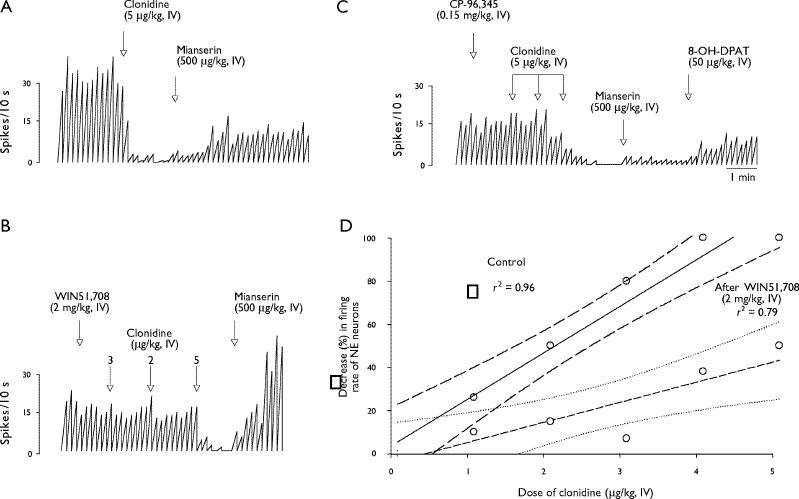
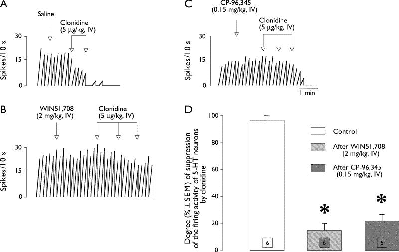
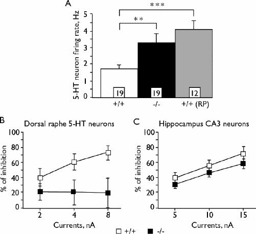
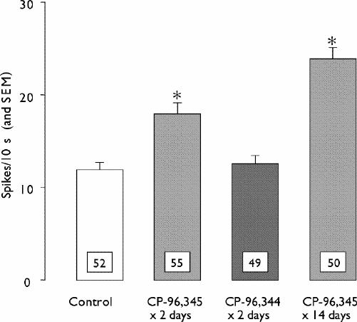
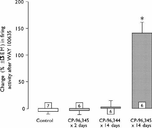
Substance P (neurokinin-1 [NK1]) receptor antagonists appear to be effective antidepressant and anxiolytic agents, as indicated in 3 double-blind clinical trials. In laboratory animals, they promptly attenuate the responsiveness of serotonin (5-hydroxytryptamine [5-HT]) and norepinephrine (NE) neurons to agonists of their cell-body autoreceptors, as is the case for some antidepressant drugs that are currently in clinical use. Long-term, but not subacute, antagonism of NK1 receptors in rats increases 5-HT transmission in the hippocampus, a property common to all antidepressant treatments tested thus far. This enhancement seems to be mediated by a time-dependent increase in the firing rate of 5-HT neurons. Mice with the NK1 receptor deleted from their genetic code also have an increased firing rate of 5-HT neurons. Taken together, these observations strongly suggest that NK1 antagonists could become a new class of antidepressant and anxiolytic agents.

Les antagonistes des récepteurs de la substance P (neurokinine-1 [NK1]) semblent efficaces comme antidépresseurs et anxiolytiques, ainsi que l'indiquent trois études cliniques à double insu. Chez des animaux de laboratoire, ces agents atténuent rapidement la réponse des neurones à sérotonine (5-hydroxytryptamine [5-HT]) et à norépinéphrine (NE) aux agonistes de leurs autorécepteurs du corps cellulaire, comme dans le cas de certains antidépresseurs actuellement utilisés en clinique. L'antagonisme à long terme mais non subaigu des récepteurs de la NK1 chez le rat accroÎt la transmission de la 5-HT dans l'hippocampe, caractéristique commune à tous les traitements aux antidépresseurs étudiés à ce jour. Une augmentation de la fréquence de décharge des neurones de la 5-HT liée au temps semble intervenir dans cette élévation. Les souris dont on a éliminé le récepteur de la NK1 du code génétique ont aussi une fréquence de décharge plus élevée des neurones de la 5-HT. Globalement, ces observations indiquent fortement que les antagonistes de la NK1 pourraient devenir une nouvelle catégorie d'antidépresseurs et d'anxiolytiques.


Figures

None
Fig. 1: Interconnections between serotonin (5-HT) and norepinephrine (NE) neurons in the brain stem and their common projection sites in the forebrain. The pathway from the locus coeruleus to the dorsal raphe is a monosynaptic projection terminating on α1-adrenergic receptors on 5-HT neurons, as depicted. The pathway from the dorsal raphe to the locus coeruleus is composed of an apposition of 3 synapses, with the excitatory 5-HT2A receptor being located on a γ-aminobutyric acid (GABA) interneuron projecting to the NE neurons of the locus coeruleus.8 At the level of the projections of 5-HT neurons, there is an α2-adrenergic receptor, not depicted on the diagram, which exerts a negative feedback action on 5-HT release. The (+) and (–) signs represent excitatory and inhibitory actions, respectively, on the function of the neurons. The firing activity of the neurons is represented by the action potential waves in red on the axons of the neurons.
None
Fig. 2: Photomicrographs of 5-HT neurons in the dorsal raphe labelled with antibodies for tryptophan hydroxylase (a and d) and for the neurokinin-1 receptors (NK1 R) (b and e). In the lowest 2 left-side images (c and f), the upper images are superimposed, showing that the NK1 immunoreactivity is not located on the 5-HT neurons themselves. The arrowhead indicates a neuron stained with the tryptophan hydroxylase antibody (PH8) and the arrow shows an NK1 receptor-positive neuron in the same area. Photomicrographs of NE neurons in the locus coeruleus labelled with antibodies for tyrosine hydroxylase (g and j) and the NK1 R (h and k). In the lowest 2 right-side images (i and l), the upper images are superimposed, showing that the immunoreactivity is located on the NE neurons themselves. Reproduced with permission from the National Academy of Sciences (Proc Natl Acad Sci U S A 2001;98:1912-7).
None
Fig. 3: Integrated firing-rate histograms of NE neurons (A–C) recorded in the locus coeruleus of anesthetized rats, showing their attenuated responsiveness to the α2-adrenergic receptor agonist clonidine after the systemic injection of the NK1 receptor antagonists. Mianserin was used as an α2-adrenergic autoreceptor antagonist to ascertain the noradrenergic nature of the neurons recorded and to show that the suppression of firing was not merely the result of losing the recording signal. In D are represented the dose-response curves for intravenous injections of 5 μg/kg of clonidine in drug-naïve rats and in rats that had received an NK1 antagonist beforehand. Each symbol represents the response of 1 neuron to the first dose of clonidine in 1 rat. The curved lines depict the standard errors of the regression lines. The correlation coefficients (r2) are given within the graph. Reproduced with permission from Lippincott Williams & Wilkins (Neuroreport 2000;11:1323-7).
None
Fig. 4: Integrated firing-rate histograms of 5-HT neurons (A–C) recorded in the dorsal raphe of anesthetized rats, showing their attenuated responsiveness to the α2-adrenergic autoreceptor agonist clonidine after the systemic injection of the NK1 receptor antagonists. In D are represented the mean responses to the first dose of clonidine (5 μg/kg) in drug-naïve rats and in rats that had received an NK1 antagonist beforehand. *p < 0.05 when compared with the control value using the Student's t test. Reproduced with permission from Lippincott Williams & Wilkins (Neuroreport 2000;11:1323-7).
None
Fig. 5A: Mean (and standard error of the mean [SEM]) firing rates of 5-HT neurons recorded in the dorsal raphe of anesthetized wild-type mice (NK1 +/+), in null mutant mice (NK1 –/–) and in wild-type mice after the systemic injection of the NK1 antagonist RP 67580. B: Decreased responsiveness of 5-HT neurons and of (C) hippocampus CA3 pyramidal neurons to the direct iontophoretic application of the 5-HT1A agonist 8-OH-DPAT, using 3 currents (nA) to eject the drug from the recording electrode, in NK1 –/– mice when compared with that assessed in wild-type mice. Reproduced with permission from the National Academy of Sciences (Proc Natl Acad Sci U S A 2001;98:1912-7).
None
Fig. 6: Histograms illustrate the mean (and SEM) firing rate of 5-HT neurons recorded in the dorsal raphe of anesthetized saline-treated rats and in anesthetized rats that received the active NK1 receptor antagonist CP-96,345 or the NK1-inactive enantiomer CP-96,344. The number of neurons recorded in each group is indicated at the bottom of each histogram. *p < 0.05 when compared with the control group. Reproduced with permission from Elsevier (Biol Psychiatry 2001;50:191-9).
None
Fig. 7: Changes in the spontaneous firing activity of hippocampus CA3 pyramidal neurons, recorded in anesthetized rats, after the intravenous injection of the 5-HT1A antagonist WAY 100,635 to determine the degree of activation of postsynaptic 5-HT1A receptors in that forebrain structure. An increase in firing rate represents a disinhibitory effect exerted by the antagonist, thereby preventing an increased inhibitory tone being mediated by increased 5-HT transmission in the hippocampus. The number of rats tested in each group is given within each histogram. The variance is expressed as the SEM. Note that only the long-term treatment with the active NK1 antagonist (CP-96,345) increased 5-HT transmission. *p < 0.05 when compared with the control group. Reproduced with permission from Elsevier (Biol Psychiatry 2001;50:191-9).

Similar articles

Cited by

References

    1. Thase ME, Entsuah AR, Rudolph RL. Remission rates during treatment with venlafaxine or selective serotonin reuptake inhibitors. Br J Psychiatry 2001;178:234-41. - PubMed
    1. Thase ME, Ninan PT. New goals in the treatment of depression: moving toward recovery. Psychopharmacol Bull 2002; 36(Suppl 2):24-35. - PubMed
    1. Kramer MS, Cutler N, Feighner J, Shrivastava R, Carman J, Sramek JJ, et al. Distinct mechanism for antidepressant activity by blockade of central substance P receptors. Science 1998; 281:1640-5. - PubMed
    1. Guyenet PG, Aghajanian GK. Excitation of neurons in the nucleus locus coeruleus by substance P and related peptides. Brain Res 1977;136:178-84. - PubMed
    1. McLean S, Ganong AH, Seeger TF, Bryce DK, Pratt KG, Reynolds LS, et al. Activity and distribution of binding sites in brain of a nonpeptide substance P (NK1) receptor antagonist. Science 1991;251:437-9. - PubMed

Publication types

MeSH terms