. 2004 Jun 15;18(12):1397-412.
doi: 10.1101/gad.301404. Epub 2004 Jun 2.

The loss of circadian PAR bZip transcription factors results in epilepsy

Affiliations

The loss of circadian PAR bZip transcription factors results in epilepsy

Frédéric Gachon et al. Genes Dev. .

Abstract

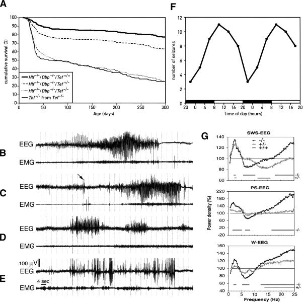
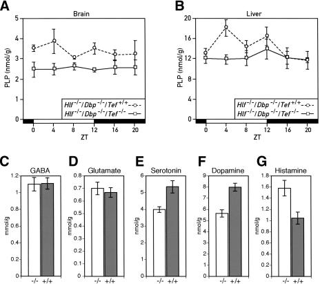
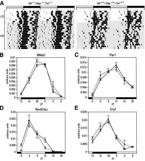
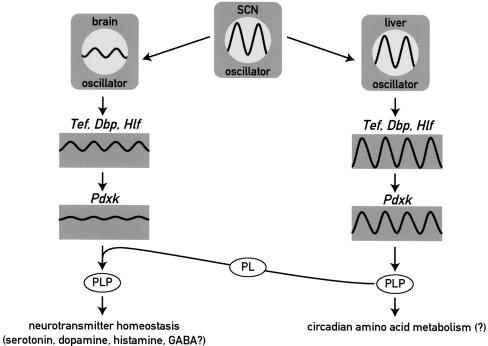
DBP (albumin D-site-binding protein), HLF (hepatic leukemia factor), and TEF (thyrotroph embryonic factor) are the three members of the PAR bZip (proline and acidic amino acid-rich basic leucine zipper) transcription factor family. All three of these transcriptional regulatory proteins accumulate with robust circadian rhythms in tissues with high amplitudes of clock gene expression, such as the suprachiasmatic nucleus (SCN) and the liver. However, they are expressed at nearly invariable levels in most brain regions, in which clock gene expression only cycles with low amplitude. Here we show that mice deficient for all three PAR bZip proteins are highly susceptible to generalized spontaneous and audiogenic epilepsies that frequently are lethal. Transcriptome profiling revealed pyridoxal kinase (Pdxk) as a target gene of PAR bZip proteins in both liver and brain. Pyridoxal kinase converts vitamin B6 derivatives into pyridoxal phosphate (PLP), the coenzyme of many enzymes involved in amino acid and neurotransmitter metabolism. PAR bZip-deficient mice show decreased brain levels of PLP, serotonin, and dopamine, and such changes have previously been reported to cause epilepsies in other systems. Hence, the expression of some clock-controlled genes, such as Pdxk, may have to remain within narrow limits in the brain. This could explain why the circadian oscillator has evolved to generate only low-amplitude cycles in most brain regions.


Figures

Figure 1.
Figure 1.
Temporal expression of mRNAs encoding PAR bZip transcription factors and clock proteins in mouse liver and brain. mRNA levels were estimated by TaqMan real-time RT–PCR and normalized to the values obtained for Gapdh mRNA. The values for Dbp, Tef, and Hlf transcripts can be directly compared, since the values were corrected for PCR efficiencies (see Materials and Methods). Mean values ± S.E.M. obtained from four animals are given in all diagrams. The Zeitgeber times (ZT) at which the animals were sacrificed are indicated on the abscissa of the diagrams. (A) Circadian expression PAR bZip transcription factor transcripts in the mouse liver. (B) Temporal expression of Dbp, Tef, and Hlf mRNAs in the brain. (C–F) Temporal expression of mPer1 (C), mPer2 (D), Rev-erbα (E), and BMal1 (F) transcripts in mouse liver and brain.
Figure 2.
Figure 2.
Disruption of the Dbp, Tef, and Hlf alleles by homologous recombination. (A) Structure of the Dbp-null allele. The entire coding sequence was replaced by a LacZNeo cassette (for details, see Lopez-Molina et al. 1997). (B) Strategy used to delete the activation domain of the Hlf allele. The cartoon displays a map of the Hlf allele, with its three promoters (α, β, and γ) and its four polyadenylation sites (vertical arrows). The positions of the recognition sites for the following restriction endonucleases are given. (E) EcoRI; (B) BamHI; (X) XbaI; (No) NotI. ATG and TAA indicate the positions of the initiation and termination codons, respectively. The structure of the targeting vector, in which part of exon 2 containing the activation domain and part of intron 2 have been replaced by a LacZNeo cassette, is given below the Hlf locus. (C) Strategy used to delete the activation domain of Tef allele. The cartoon displays a map of the Tef allele, with its two promoters (α and β). The positions of the recognition sites for the following restriction endonucleases are given. (N) NcoI; (X) XbaI; (No) NotI. ATG and TAA indicate the positions of the initiation and termination codons, respectively. The structure of the targeting vector, in which part of exon 2 containing the activation domain and part of intron 2 have been replaced by a LacZNeo cassette, is given below the Tef locus. (D,E) Expression of the disrupted Hlf allele in mouse liver (D) and brain (E). The levels of liver Hlf and HlfΔAD transcripts were estimated by TaqMan real-time RT–PCR as in Figure 1A. The results represent means ± S.E.M. from four animals. (F,G) Expression of the mutated Tef allele in mouse liver (F) and brain (G). The levels of liver Tef and TefΔAD were estimated by TaqMan real-time RT–PCR as in Figure 1A. The results represent means ± S.E.M. from four animals.
Figure 3.
Figure 3.
Phenotypes of the Hlf/Dbp/Tef triple-knockout mice. (A) Increased mortality in young triple-knockout mice. About 50% of homozygous triple-knockout mice (Hlf-/-/Dbp-/-/Tef-/-) died between 20 and 40 d of age, irrespective of whether these animals were the offspring of Tef heterozygous, double-knockout mice (Hlf-/-/Dbp-/-/Tef+/-) or homozygous triple-knockout parents (Hlf-/-/Dbp-/-/Tef-/-, labeled as Tef-/- in the figure). After this age, triple-knockout animals continued to die progressively and had an expected life span inferior to that of double-knockout animals. All double-knockout mice (Hlf-/-/Dbp-/-) and Tef heterozygous, double-knockout mice (Hlf-/-/Dbp-/-/Tef+/-) were the offspring from Tef heterozygous, double-knockout mice (Hlf-/-/Dbp-/-/Tef+/-). (B,C) Spontaneous epileptic seizure in triple-knockout animals. All three genotypes were implanted with chronic EEG and EMG electrodes to characterize the seizure activity. Spontaneous tonic–clonic seizures only occurred in triple KO mice and primarily during slow-wave sleep (SWS). The arrow in the EEG from the individual presented in C points to an abrupt sharp wave (myoclonus) followed by a generalized epileptic seizure. (D) Absence seizure in a Tef heterozygous, double-knockout animal. Several absence-like seizures without any sign of EEG hypoactivity were also recorded in heterozygous mice. These occurred during SWS as well, particularly at the transition from waking to sleep. (E) Epileptic EEG activity in a Tef heterozygous, double-knockout animal. The most common form of epileptic activity in such mice was characterized by high-voltage sharp waves, similar to those observed in homozygous triple-knockout mice before generalized seizures initiated. We never recorded any generalized seizures in mice containing at least one Tef wild-type allele. (F) Temporal occurrence of seizures. The cartoon represents the occurrence of seizure in triple-knockout mice during 4-h intervals during the day. The lights were turned on and off at 8 a.m. and 8 p.m., respectively. For esthetic reasons the values were double-plotted. (G) EEG spectral analysis of homozygous and Tef heterozygous, double-knockout mice. The sleep EEG was recorded during a 12-h daytime period in nine triple, six heterozygous, and six double-knockout mice. Spectral analysis was performed on all 4-sec epochs without any seizure activity. Mean spectral profiles for each vigilance state were calculated over each genotype and expressed as a percentage of the mean values in double-knockout mice. Horizontal lines on the bottom of each panel connect those frequency bins in which power density is significantly different from that of double-knockout mice (indicated as 100% throughout the frequency range).
Figure 4.
Figure 4.
The transcription of the pyridoxal kinase gene (Pdxk) is regulated by PAR bZip transcription factors. (A) Sequence comparison of the 5′ proximal region of the Pdxk genes from mouse, rat, and man. The transcription initiation sites, indicated by bent arrows, have been mapped on the mouse genomic sequence by RACE analysis. Three RACE products have been sequenced for each of the two start sites represented by more prominent arrows. Start sites represented by single RACE products are depicted by small arrows. A PAR bZIP transcription factor response element (PARRE) is located within the 5′-moiety of the first intron. The PARRE consensus sequence is given below the element (R = A or G, Y = C or T). (B) Binding of liver nuclear proteins to the intronic PARRE. Electomobility shift assays (EMSA) with a radio-labeled oligonucleotide encompassing the PARRE and liver nuclear proteins from triple-knockout mice, double-knockout mice and wild-type animals harvested at 4-h intervals around the clock. ZT stands for Zeitgeber time. The lights were turned on and off at ZT0 and ZT12, respectively. (C) Binding of brain nuclear proteins to the Pdxk PARRE. EMSA were carried out as in B, but with brain nuclear proteins. (D) Temporal expression of Pdxk RNA in the liver of double-knockout mice and triple-knockout mice. The relative accumulation of Pdxk mRNA was determined by TaqMan real-time RT–PCR with liver whole-cell RNAs prepared from animals sacrificed at the indicated Zeitgeber times (ZT). Mean values ± S.E.M. from four animals are shown. ANOVA revealed significant circadian rhythmicity only for the hepatic Pdxk mRNA expression of double-knockout animals (F[5,18] = 7.73, p < 0.0005). The difference between genotypes is highly significant (ANOVA F[1,46] = 32.03, p < 1.10-6). (E) Temporal expression of Pdxk mRNA in the brain of double-knockout and triple-knockout mice. The relative levels of Pdxk mRNA were determined as in D. The values are means ± S.E.M. from four animals. Although the transcript levels of double-knockout mice measured at ZT8 appear somewhat lower than those measured at other ZTs, ANOVA did not reveal a circadian Pdxk expression. The difference between genotype is highly significant (ANOVA F[1,46] = 41.61, p < 1.10-7). (F) Expression of Pdxk RNA in different regions of the brain. Coronal brain sections were prepared from double- or triple-knockout mice sacrificed at ZT10 and hybridized to a 35S-labeled antisense (top and middle panels) or a sense (bottom panel) Pdxk RNA probe. (CP) Caudate putamen (striatum); (LS) lateral septal nucleus; (MH) medial habenula; (HF) hippocampal formation; (BMA) baso-medial amygdaloid nucleus; (T) thalamus; (ZI) zona incerta; (DMH) dorsomedial hypothalamic nucleus; (IC) inferior colliculus; (PAG) peri-aqueductal gray; (GLC) granular layer of cerebellum; (CGP) central gray of the pons.
Figure 5.
Figure 5.
Down-regulation of pyridoxal phosphate (PLP) levels and dysregulation of neurotransmitter metabolism in PAR bZip triple-knockout mice. (A) Temporal concentration of PLP in the brains of double- and triple-knockout mice. The PLP levels were measured by high-performance liquid chromatography (HPLC) of perchloric acid brain extracts prepared from animals sacrificed at the indicated Zeitgeber times (ZT). The values are means ± S.E.M. from three animals. The difference between the two genotypes is highly significant (ANOVA F[1,32] = 20.18, p < 1.10-4). (B) Temporal accumulation of PLP in the livers of double- and triple-knockout mice. PLP levels were determined as in A, and the values are means ± S.E.M. from at least six animals. ANOVA revealed significant circadian rhythmicity in PLP levels in double-knockout animals (F[5,32] = 2.60, p < 0.05). The difference between geno-types is also statistically significant (ANOVA F[1,73] = 5.31, p < 0.025). (C,D) GABA (C) and Glutamate (D) concentration in the brains of Hlf-/-/Dbp-/-/Tef-/- (-/-) and Hlf-/-/Dbp-/-/Tef+/+ (+/+) mice. No statistically significant difference could be found between the two genotypes. The values are means ± S.E.M. from 12 animals of each genotype. (E,F,G) Concentration of serotonin (E), dopamine (F) and histamine (G) in the brains of Hlf-/-/Dbp-/-/Tef-/- (-/-) and Hlf-/-/Dbp-/-/Tef+/+ (+/+) mice. The difference between genotype is statistically significant (Student's t test: p < 0.005 for serotonin, p < 0.0001 for dopamine and p < 0.01 for histamine). The values are means ± S.E.M. from 12 animals of each genotype.
Figure 6.
Figure 6.
Circadian behavior and gene expression in PAR bZip triple-knockout mice. (A) Circadian locomotor (wheel running) activity of Hlf/Dbp/Tef triple-knockout (left panel) and wild-type (right panel) mice. In each actogram, the first few days were recorded under LD conditions (lights on at 7 a.m.; lights off at 7 p.m.). Time spans during which the light was switched off are marked by gray shadowing. The free-running periods in constant darkness of these mice show no difference between geno-types (Hlf-/-/Dbp-/-/Tef-/-: 23.63 ± 0.33 h; wild-type: 23.68 ± 0.33 h). (BE) Temporal expression of BMal1 (B), mPer1 (C), Rev-erbα (D), and Cry1 (E) transcripts in liver, as determined by real-time RT–PCR in Hlf/Dbp/Tef triple-knockout (solid line) and wild-type mice (dotted line). Mean values ± S.E.M. obtained from four animals are given in all diagrams. The Zeitgeber times (ZT) at which the animals were sacrificed are indicated on the abscissa of the diagrams.
Figure 7.
Figure 7.
Model showing the regulation and function of Pdxk in brain and liver. The molecular circadian oscillator generates high amplitude cycles and low amplitude cycles of PAR bZip gene expression in liver and brain, respectively. Accordingly, target genes of PAR bZip transcription factors, such as Pdxk, are expressed in a strongly circadian manner in liver and at nearly constant levels in the brain. As PLP, the product of the PDXK reaction is a coenzyme for amino acid decarboxylases and aminotransferases, the rhythmic production of PLP in the liver may contribute to circadian amino acid metabolism. In the brain, nearly invariable Pdxk expression may be essential for regulating neurotransmitter homeostasis, as moderate decreases in PLP levels may result in neurotransmitter deficiencies and epileptic attacks (see text). (SCN) suprachiasmatic nucleus harboring the central circadian pacemaker; (PLP) pyridoxal phosphate; (PL) pyridoxal.

References

    1. Babity J.M., Armstrong, J.N., Plumier, J.C., Currie, R.W., and Robertson, H.A. 1997. A novel seizure-induced synaptotagmin gene identified by differential display. Proc. Natl. Acad. Sci. 94: 2638-2641. - PMC - PubMed
    1. Battaglioli G., Liu, H., and Martin, D.L. 2003. Kinetic differences between the isoforms of glutamate decarboxylase: Implications for the regulation of GABA synthesis. J. Neurochem. 86: 879-887. - PubMed
    1. Bell R.R. and Haskell, B.E. 1971. Metabolism of vitamin B 6 in the I-strain mouse. I. Absorption, excretion, and conversion of vitamin to enzyme co-factor. Arch. Biochem. Biophys. 147: 588-601. - PubMed
    1. Bell R.R., Blanchard, C.A., and Haskell, B.E. 1971. Metabolism of vitamin B6 in the I-strain mouse. II. Oxidation of pyridoxal. Arch. Biochem. Biophys. 147: 602-611. - PubMed
    1. Blaschko H. and Buffoni, F. 1965. Pyridoxal phosphate as a constituent of the histaminase (benzylamine oxidase) of pig plasma. Proc. R. Soc. Lond. B. Biol. Sci. 163: 45-60. - PubMed

Publication types

MeSH terms