. 2004 Jul;131(13):3147-56.
doi: 10.1242/dev.01187. Epub 2004 Jun 2.

HUA ENHANCER3 reveals a role for a cyclin-dependent protein kinase in the specification of floral organ identity in Arabidopsis

Affiliations

HUA ENHANCER3 reveals a role for a cyclin-dependent protein kinase in the specification of floral organ identity in Arabidopsis

Wenming Wang et al. Development. 2004 Jul.

Abstract

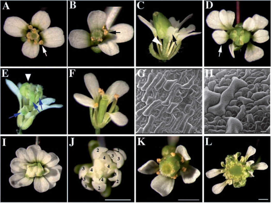
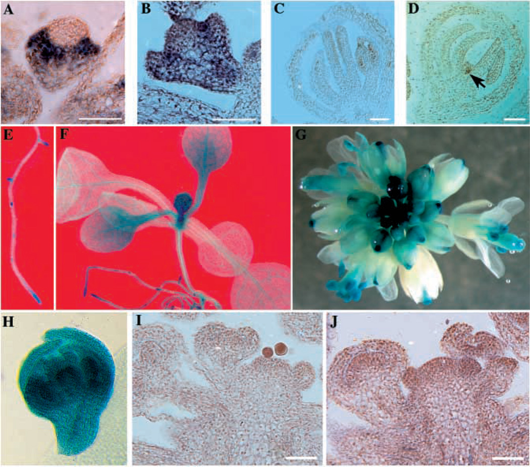
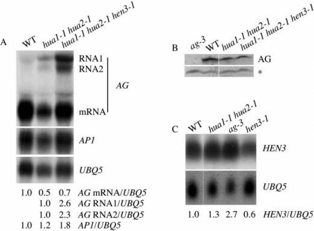
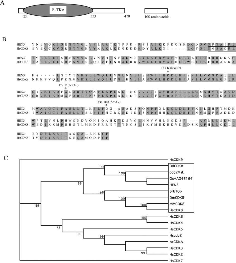
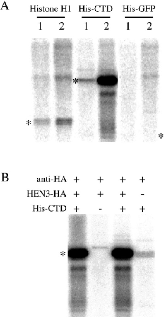
In plants, organs are generated post-embryonically from highly organized structures known as meristems. Cell division in the meristem is closely integrated with cell fate specification and organ formation. The presence of multiple cyclin-dependent kinases (CDKs) and their partner cyclins in plants and other multicellular organisms probably reflects the complexity of cell cycle regulation within developmental contexts. The Arabidopsis genome encodes at least eight CDKs and 30 cyclins. However, no mutants in any CDKs have been reported, and the function of the great majority of these genes in plant development is unknown. We show that HUA ENHANCER3 (HEN3), which encodes CDKE, a homolog of mammalian CDK8, is required for the specification of stamen and carpel identities and for the proper termination of stem cells in the floral meristem. Therefore, CDK8 plays a role in cell differentiation in a multicellular organism.


Figures

Fig. 1
Fig. 1
Floral phenotypes caused by hen3 mutations. (A) A wild-type flower with stamens in the third whorl (arrow) and carpels in the fourth whorl. (B) A hua1-1 hua2-1 flower with stamens in the third whorl (arrow) and carpels in the fourth whorl. (C) A hua1-1 hua2-1 hen3-1 flower with petals in the third whorl (arrow) and an enlarged gynoecium with a gynophore at the base (arrowhead), which indicates that the gynoecium has partial floral character. (D) A hua1-1 hua2-1 hen3-2 flower with petals in the third whorl (arrow) and an enlarged gynoecium in the center. (E) A hua1-1 hua2-1 hen3-3 flower with an additional flower in the center (arrowhead). The blue arrows indicate three unfused carpels. (F) A hen3-1 flower with normal organ identity. (G) Epidermal valve cells with a smooth surface from the bottom of a hua1-1 hua2-1 ovary. (H) Epidermal valve cells from a hua1-1 hua2-1 hen3-1 ovary. The cells have epicuticular striations similar to epidermal cells on sepals. (I) A hua1-1 hua2-1 hen3-1 ag-1 flower. (J) A hen3-1 ag-1 flower. The numbers indicate the floral whorls. The fourth whorl sepals are largely transformed to petals. (K) A clv1-4 flower. (L) A hua1-1 hua2-1 hen3-1 clv1-4 flower, with a massive amount of undifferentiated cells in the center. Scale bars: 10 µm in G,H; 1 mm in J–L.
Fig. 2
Fig. 2
Expression patterns of AP1, WUS, and HEN3 in plants. (A,B) In situ hybridization on sections of stage 3 flowers to detect AP1 RNA. (A) A hua1-1 hua2-1 flower. (B) A hua1-1 hua2-1 hen3-1 flower. AP1 RNA is found throughout the flower in hua1-1 hua2-1 hen3-1. (C,D) In situ hybridization on sections of stage 9 flowers to detect WUS RNA. (C) A stage 9 hua1-1 hua2-1 flower that has ceased to express WUS. (D) A stage 9 hua1-1 hua2-1 hen3-1 flower that still expresses WUS (arrow). (E–H) GUS staining of HEN3-GUS transgenic lines. HEN3-GUS is found in proliferating tissues or organs. (E) HEN3-GUS is present at the root tip and lateral root primordia. (F) HEN3-GUS is present at the shoot tip, in young leaves, and at the base of growing leaves. (G) HEN3-GUS is present in young flowers. (H) HEN3-GUS is found throughout a stage 7 flower. (I,J) In situ hybridization on longitudinal sections of wild-type inflorescences using either the sense (I) or the antisense (J) HEN3 probe. HEN3 RNA is found at a low level throughout the inflorescence meristem and the floral meristems. Scale bars: 50 µm.
Fig. 3
Fig. 3
AP1, AG and HEN3 expression in various genotypes. (A) RNA filter hybridization to examine AG and AP1 RNA levels in various genotypes as indicated. The two AG RNAs containing 2nd intron sequences are marked as RNA1 and RNA2. The abundance of AP1 and AG RNAs is indicated by the numbers below the gel images. (B) Western blotting on total proteins from inflorescences from various genotypes using anti-AG antisera. ag-3 serves as a negative control. AG protein abundance is similar in hua1-1 hua2-1 and hua1-1 hua2-1 hen3-1 flowers. The band marked by an asterisk is a crossreacting, non-AG species from the same blot. (C) RNA filter hybridization to determine the abundance of HEN3 RNA from inflorescences of various genotypes. The abundance relative to wild type is indicated by numbers below the gel images.
Fig. 4
Fig. 4
HEN3 is closely related to CDK8. (A) A diagram of HEN3 protein with the serine-threonine kinase domain represented by the oval. (B) A Clustal W alignment of the kinase domains in HEN3 and human CDK8. Identical and similar amino acids are shaded. The amino acid positions and nature of the three hen3 alleles are indicated above the sequences. The cyclin-binding motifs of HEN3 and CDK8 are underlined. (C) A bootstrap consensus phylogenetic tree involving nine different human CDKs, CDK8 from Drosophila (DmCDK8), mouse (MmCDK8), Dictyostelium (DdCDK8) and yeast (Srb10p), three E type CDKs from plants, and the Arabidopsis CDKA (AtCDKA). Cdc2MsE and OsAAG46164 are CDKEs from alfalfa and rice, respectively. The tree was constructed using the neighbor joining method and the bootstrap consensus was generated from 1000 replications. The numbers represent the percentage occurrence of the nodes in the replications. Only the protein kinase domains from these proteins were used in the analysis. The CDK8 clade is indicated by the rectangle.
Fig. 5
Fig. 5
HEN3 has CTD kinase activity. HEN3-HA transgenic plants or control non-transgenic plants were immunoprecipitated with an anti-HA monoclonal antibody, and the immunoprecipitate was used in a kinase assay with 10 µg purified histone H1, 6×His-CTD or 6×His-GFP as substrates. The positions of the substrate proteins are illustrated by the asterisks. The amount of substrate proteins is identical in each lane. (A) The anti-HA (lanes 2) but not the anti-protein C (lanes 1) immunoprecipitate from HEN3-HA transgenic plants displays kinase activity on 6×His-CTD. Histone H1 and 6×His-GFP are not phosphorylated by the anti-HA immunoprecipitate. (B) The anti-HA immunoprecipitate from HEN3-HA transgenic plants but not from wild-type plants phosphorylates 6×His-CTD.
Fig. 6
Fig. 6
Effects of hen3 mutations on leaf size and leaf epidermal cell size. (A–C) Dissected rosette leaves 1–7 (arranged from left to right) from hen3 (top) and wild-type (bottom) plants that were grown side by side. (A) hen3-1 and wild type. (B) hen3-2 and wild type. (C) hen3-3 and wild type. Note that the first two leaves are not affected as much as the later leaves by the hen3 mutations. The hen3 mutations all lead to a reduction in leaf size. (D) Area (cm2) of the fully expanded fifth leaf from wild-type and hen3 genotypes. Ten leaves were measured for each genotype. (E) Surface area (µm2) of adaxial epidermal cells on the fifth leaf of wild-type and hen3 genotypes. The number of cells measured was 95, 141, 173 and 143 for wild type, hen3-1, hen3-2 and hen3-3, respectively. The large standard error is largely due to the intrinsic variation in cell size on the leaf epidermis. (F) Number of epidermal pavement cells in a 70,000 µm2 area from similar positions in the fifth leaves of various genotypes. Measurements were performed on SEM images of three leaves from each genotype. The error bars in D–F represent standard errors.

Similar articles

Cited by

References

    1. Akoulitchev S, Chuikov S, Reinberg D. TFIIH is negatively regulated by cdk8-containing mediator complexes. Nature. 2000;407:102–106. - PubMed
    1. Barroco RM, de Veylder L, Magyar Z, Engler G, Inze D, Mironov V. Novel complexes of cyclin-dependent kinases and a cyclin-like protein from Arabidopsis thaliana with a function unrelated to cell division. Cell. Mol. Life Sci. 2003;60:401–412. - PMC - PubMed
    1. Boniotti MB, Gutierrez C. A cell-cycle-regulated kinase activity phosphorylates plant retinoblastoma protein and contains, in Arabidopsis, a CDKA/cyclin D complex. Plant J. 2001;28:341–350. - PubMed
    1. Bowman JL, Smyth DR, Meyerowitz EM. Genes directing flower development in Arabidopsis. Plant Cell. 1989;1:37–52. - PMC - PubMed
    1. Chen X. A microRNA as a translational repressor of APETALA2 in Arabidopsis flower development. Science. 2004;303:2022–2025. - PMC - PubMed

Publication types

MeSH terms