. 2004 Jun 1;18(11):1345-58.
doi: 10.1101/gad.1184904.

Signaling hierarchy downstream of retinoic acid that independently regulates vascular remodeling and endothelial cell proliferation

Affiliations

Signaling hierarchy downstream of retinoic acid that independently regulates vascular remodeling and endothelial cell proliferation

Brenda L Bohnsack et al. Genes Dev. .

Abstract

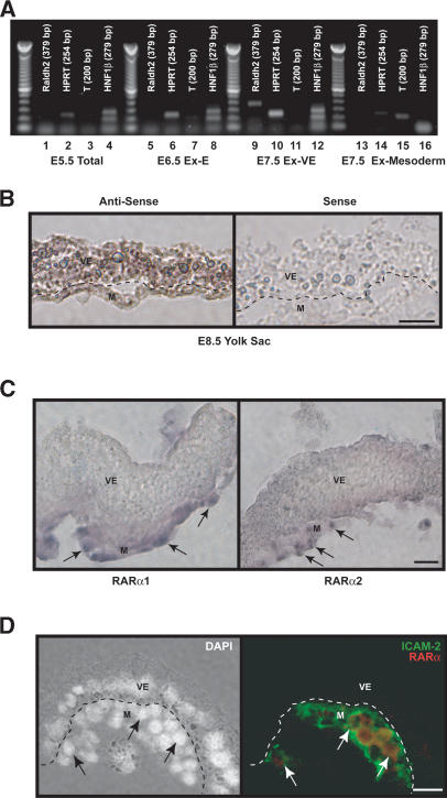
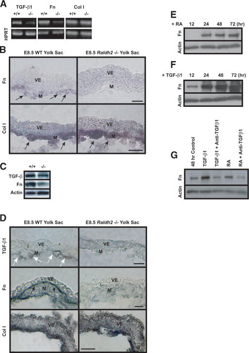
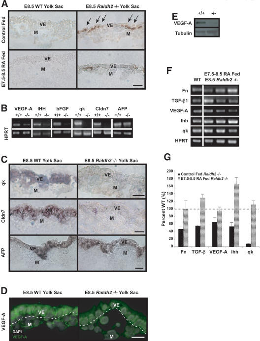
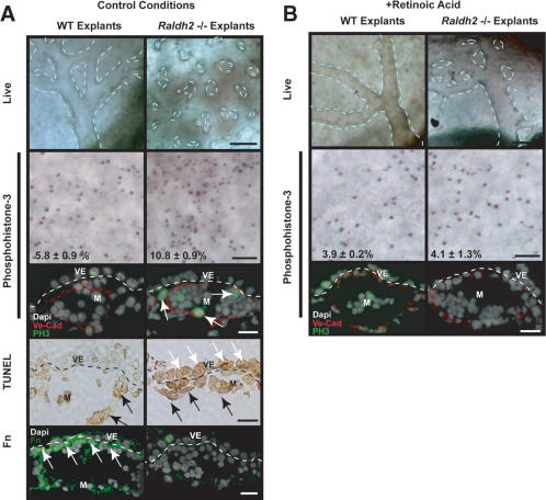
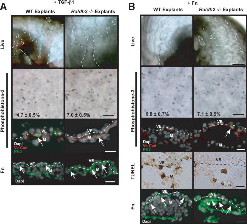
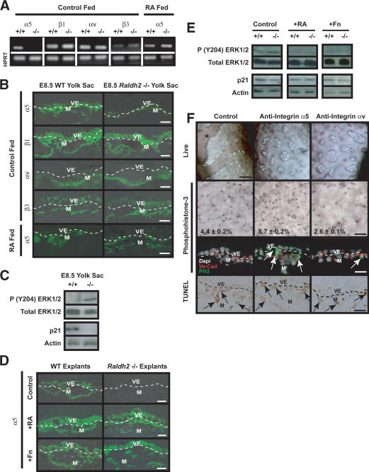
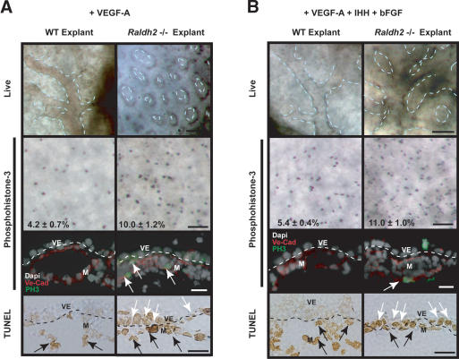
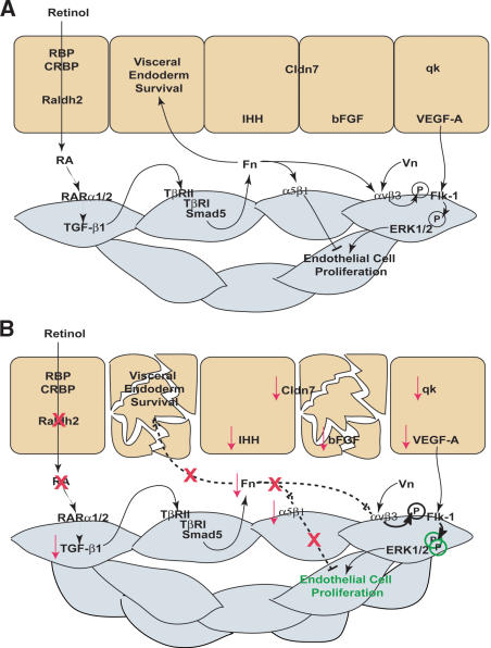
We previously demonstrated that during vascular morphogenesis, retinoic acid (RA) is required for the control of endothelial cell proliferation and capillary plexus remodeling. Herein, we investigate the mechanisms by which RA regulates these processes in the yolk sac. We found that although the enzyme required for RA production during early embryogenesis, retinaldehyde dehydrogenase-2 (Raldh2), was expressed in the visceral endoderm, RA receptors alpha1 and alpha2 were expressed in endothelial cells in the mesoderm, indicating that they are direct targets of RA. In Raldh2(-/-) embryos, there was down-regulation of TGF-beta1, fibronectin (Fn) and integrin alpha5, which was associated with decreased visceral endoderm survival and production of VEGF-A, Indian hedgehog (IHH), and bFGF. Exogenous provision of RA or Fn to Raldh2(-/-) explants in whole mouse embryo culture restored vascular remodeling, visceral endoderm survival, as well as integrin alpha5 expression and its downstream signaling that controls endothelial growth. Exogenous provision of visceral endoderm-derived factors (VEGF-A, IHH, and bFGF) failed to rescue endothelial cell proliferative control but collectively promoted vascular remodeling, suggesting that these processes are independently regulated via a signaling hierarchy downstream of RA.


Figures

Figure 1.
Figure 1.
RA synthesis and signaling in the yolk sac. (A) Raldh2 gene expression was not detected by sqRT-PCR in RNA isolated from total E5.5 embryos (lanes 1-4) and the extraembryonic region of E6.5 embryos (lanes 5-8). RNA was also independently isolated from E7.5 visceral endoderm (lanes 9-12) and mesoderm (lanes 13-16). Raldh2 transcript was most prevalent in visceral endoderm at E7.5. HNF1β and T-brachyury (T) served as markers for visceral endoderm and mesoderm, respectively, and HPRT was used as an internal control. (B) In situ hybridization demonstrated that in E8.5 embryos, Raldh2 was most highly expressed in visceral endoderm, but is also in some endothelial cells. Bar, 25 μm. (C) In situ hybridization localized RARα1 and α2 transcripts within the mesoderm (arrows) of E8.5 yolk sac. Bar, 25 μm. (D) Immunofluorescence was used to colocalize RARα proteins with ICAM-2, a marker for endothelial cells (arrows) in E8.5 yolk sac. Bar, 25 μm. (M) Mesoderm; (V) visceral endoderm.
Figure 2.
Figure 2.
RA regulation of TGF-β1 and Fn in endothelial cells. (A) sqRT-PCR revealed decreased expression of TGF-β1 and Fn transcripts in E8.5 Raldh2-/- yolk sacs compared with wild type (WT). Col I transcript levels were similar in both Raldh2-/- and wild-type yolk sacs; HPRT was used as an internal control. (B) In situ hybridization of E8.5 yolk sac sections revealed that Fn (top) and Col I (bottom) transcripts were expressed in the mesoderm (arrows); however, Fn transcript levels were down-regulated in Raldh2-/- (right) compared with wild type (WT; left). Bar, 25 μm. (C) Western blot analyses of E8.5 Raldh2-/- mutant and wild-type (WT) yolk sacs demonstrated decreased levels of TGF-β1 and Fn protein. Actin was used as a loading control. (D) Immunohistochemistry of E8.5 yolk sac sections revealed that levels of TGF-β1 protein (top row), specifically expressed in endothelial cells (arrows), and Fn protein (middle row), deposited between the visceral endoderm and mesoderm (black arrows), were decreased in Raldh2-/- yolk sac (right) compared with wild type (WT; left). Protein levels of Col I (bottom row) were similar in Raldh2-/- and wild type. Bar, 25 μm. (E-G) Bovine aortic endothelial cells were cultured for up to 72 h in the presence of 1 μM RA (D) or 1 ng/mL TGF-β1 (F). Western blot demonstrated that RA increased Fn production over control levels at 24 h (E,G). TGF-β1 significantly increased Fn production in endothelial cells starting at 12 h (F,G). (G) Neutralizing anti-sera against TGF-β1, TGF-β2, and TGF-β3 suppressed RA-induced Fn production in endothelial cells, as well as TGF-β1-induced Fn production. (M) Mesoderm; (V) visceral endoderm.
Figure 3.
Figure 3.
RA maintains visceral endoderm integrity and differentiated function. (A) TUNEL assay revealed a 15-fold increase of apoptotic cells localized to extraembryonic visceral endoderm in control (top) E8.5 Raldh2-/- yolk sacs (right) compared with wild type (WT; left). Feeding RA to pregnant mothers from E7.5-E8.5 decreased visceral endoderm apoptosis by 11-fold in E8.5 Raldh2-/- yolk sacs (bottom). Bar, 25 μm. (B) sqRT-PCR demonstrated that in E8.5 yolk sacs, levels of VEGF-A164, IHH, bFGF, qk, and Claudin 7 (Cldn7) were down-regulated in Raldh2-/- versus wild type (WT); AFP was similar in Raldh2-/- and wild type. HPRT was used as an internal control. (C) In situ hybridization of E8.5 yolk sac sections for qk (top row), and Cldn7 (middle row) localized these transcripts to extraembryonic visceral endoderm where they were down-regulated in Raldh2-/- (middle). (Bottom row) AFP transcripts were localized to the embryonic visceral endoderm and were similar in Raldh2-/- and wild type. Bar, 25 μm. (D,E) Immunofluorescence and Western blot analyses for VEGF-A protein revealed decreased levels in E8.5 Raldh2-/- mutant yolk sacs compared with wild type (WT). Tubulin was used as a loading control. (F) Feeding pregnant mothers RA from E7.5-E8.5 restored transcript levels of TGF-β1, Fn, VEGF-A164, IHH, and qk in three individual E8.5 Raldh2-/- yolk sacs to wild-type (WT) levels as determined by sqRT-PCR. (G) sqRT-PCR of RNA from control (black bars) and RA fed (striped bars) E8.5 Raldh2-/- yolk sacs, represented as percent of similarly fed wild-type littermates, demonstrated that RA restored levels of transcripts down-regulated in control-treated Raldh2-/- yolk sacs. n = 4-5. (M) Mesoderm; (V) visceral endoderm.
Figure 4.
Figure 4.
In vitro Raldh2-/- yolk sac vascular phenotype and rescue with RA. (A) Wild-type (WT; left) and Raldh2-/- (right) explants after 48 h in control conditions in whole embryo culture recapitulated our in vivo findings as Raldh2-/- yolk sac failed to remodel (top row; bar, 100 μm). Dashed lines highlight vascular patterning in yolk sac. Whole-mount immunostaining of yolk sac tissues (second row; bar, 100 μm) with anti-phosphohistone-3 demonstrated a twofold increase in the mitotic index of endothelial cells in Raldh2-/- explants compared with wild type (WT), also corroborating in vivo findings. Furthermore, immunofluorescence (third row; bar, 25 μm) to colocalize phosphohistone-3 (green) with VE-Cad (red) demonstrated that the proliferating cells were predominantly endothelium (arrows). TUNEL assay of yolk sac sections (fourth row; bar, 25 μm) revealed a 10-fold increase in apoptosis in extraembryonic visceral endoderm (VE, white arrows) of Raldh2-/- explants. The level of apoptosis in mesoderm (M, black arrows) of Raldh2-/- explants was similar to that of wild type. Detection of Fn protein by immunofluorescence (bottom row; bar, 25 μm) demonstrated decreased Fn deposition specifically between the visceral endoderm and mesoderm layers (arrows) in Raldh2-/- explants. (B) Addition of 3 ng/mL RA to culture media restored vascular remodeling in yolk sacs of Raldh2-/- explants (top) and had no effect on wild-type development. Whole-mount (middle row; bar, 100 μm) immunostaining with antiphosphohistone-3 and immunofluorescence (bottom row; bar 25 μm) with antiphosphohistone-3 (green) and VE-cad (red) revealed that RA restored endothelial cell cycle control in Raldh2-/- explants to wild-type level.
Figure 5.
Figure 5.
TGF-β1 and Fn rescue Raldh2-/- vascular defects in vitro. (A) Treatment of Raldh2-/- and wild-type (WT) embryo explants with 10 ng/mL TGF-β1 for 48 h initiated vascular remodeling in Raldh2-/- explants (top row). Detection of proliferating cells by whole-mount (middle row; bar 100 μm) immunostaining of yolk sacs with antiphosphohistone-3 and immunofluorescence (bottom row; bar 25 μm) with antiphosphohistone-3 (green) and VE-Cad (red) revealed that TGF-β1 decreased endothelial cell proliferation in Raldh2-/- explants but not to wild-type levels. Treatment with TGF-β1 also restored Fn deposition between the visceral endoderm (VE) and mesoderm (M) as demonstrated by immunofluorescence. (B) Treatment with 100 μg/mL soluble Fn rescued vascular remodeling in yolk sac of Raldh2-/- explants (top row; bar, 100 μm). Whole-mount immunostaining of yolk sac tissues (second row; bar, 100 μm) with antiphosphohistone-3 and immunofluorescence of yolk sac sections (third row; bar, 25 μm) with anti-phosphohistone-3 (green) and VE-Cad (red) revealed that Fn rescued endothelial cell cycle control in Raldh2-/- explants to that of wild-type levels. TUNEL assay (fourth row; bar, 25 μm) demonstrated that Fn reduced the level of apoptosis in visceral endoderm of Raldh2-/- explant yolk sacs to wild-type levels and the remaining apoptotic cells in Raldh2-/- yolk sacs were in the mesoderm (arrows) similar to wild type. (Bottom row) Detection of Fn protein by immunofluorescence of yolk sac sections demonstrated that treatment of Raldh2-/- with soluble Fn restored Fn deposition between the visceral endoderm and mesoderm layers of the yolk sac (arrows).
Figure 6.
Figure 6.
Endothelial cell proliferative control is regulated by extracellular matrix proteins via integrin α5 and integrin αv. (A) sqRT-PCR analysis of E8.5 Raldh2-/- and wild-type (WT) yolk sacs revealed down-regulation of integrin α5 transcript, but no change in β1, αv, or β3 levels. Feeding RA to pregnant females from E7.5-E8.5 restored α5 transcript levels in Raldh2-/- yolk sacs. HPRT was used as an internal control. (B) Immunofluorescence demonstrated that integrin α5 protein is decreased in control fed E8.5 Raldh2-/- yolk sacs compared with wild type (WT; top row), but protein expression was restored by feeding RA to pregnant females from E7.5-E8.5 (bottom row). There were no differences in protein levels of β1 (second row), αv (third row), or β3 (fourth row) in E8.5 Raldh2-/- yolk sacs compared with wild type. (C) Western blot analyses of E8.5 Raldh2-/- and wild-type (WT) yolk sac protein demonstrated increased phosphorylation of ERK1/2 at Tyr 204 (top row) in mutant yolk sacs compared with wild type. Conversely, protein levels of p21 (third row) were decreased in Raldh2-/- yolk sacs. Total ERK (second row), and actin (bottom row) were used as loading controls. (D) Immunofluorescence of embryo culture sections revealed that integrin α5 protein, which was decreased in control treated Raldh2-/- explants (top row), was restored by treatment with 3 ng/mL RA (middle row) and 100 μg/mL Fn (bottom row). (E) Western blot analyses of yolk sacs from embryo culture explants demonstrated that phosphorylation of ERK1/2 (Tyr 204), which were increased in untreated Raldh2-/- mutants, were restored to wild-type levels by treatment with 3 ng/mL RA and 100 μg/mL Fn. Protein levels of p21 were decreased in untreated Raldh2-/- explants but restored by treatment with RA. (F) Blocking antibodies to integrin α5 (middle column) or integrin αv (right column) inhibited vascular remodeling in 40% of wild-type explants in whole embryo culture (top row; bar, 100 μm). Whole-mount immunostaining (second row; bar, 100 μm) with antiphosphohistone-3 and immunofluorescence (third row; bar, 25 μm) with antiphosphohistone-3 (green) and VE-Cad (red) revealed that blocking integrin α5 signaling increased endothelial cell proliferation. Conversely, inhibiting integrin αv resulted in decreased proliferation. TUNEL assay (bottom row) demonstrated no differences in apoptosis in the mesoderm (arrows) and visceral endoderm in control and α5 or αv antibody treated explants.
Figure 7.
Figure 7.
Visceral endoderm factors are required for vascular remodeling, but not endothelial proliferative control. (A) Treatment with 50 ng/mL VEGF-A165 in whole embryo culture failed to induce vascular remodeling in Raldh2-/- explant yolk sacs (top row; bar, 100 μm). Whole-mount immunostaining of yolk sac tissues with antiphosphohistone-3 (second row; bar, 100 μm) and immunofluorescence of yolk sac sections (third row; bar, 25 μm) with antiphosphohistone-3 (green) and VE-Cad (red) demonstrated that Raldh2-/- explants treated with VEGF-A165, had a twofold increase in proliferating endothelial cells (arrows) compared with wild type (WT). TUNEL analyses of yolk sac sections (bottom row; bar, 25 μm) revealed that VEGF-A165 treated Raldh2-/- explants had a 10-fold increase in apoptosis in visceral endoderm (VE, white arrows) but no change in the mesoderm (M, black arrows). (B) Treatment with 50 ng/mL VEGF-A165 + 1 μg/mL IHH + 100 ng/mL bFGF induced vascular remodeling in Raldh2-/- explant yolk sac (top row; bar, 100 μm). Whole-mount immunostaining of yolk sac tissues with antiphosphohistone-3 (second row; bar, 100 μm) and immunofluorescence with antiphosphohistone-3 (green) and VE-Cad (red; third row; bar, 25 μm) revealed Raldh2-/- explants had a twofold increase in endothelial cell proliferation compared with wild type. TUNEL assay of yolk sac sections (bottom row; bar, 25 μm) demonstrated that Raldh2-/- explants treated with 50 ng/mL rh-VEGF-A165 + 1 μg/mL IHH + 100 ng/mL bFGF had a 10-fold increase in apoptosis in visceral endoderm (white arrows), similar to untreated Raldh2-/- explants.
Figure 8.
Figure 8.
RA regulation of reciprocal interactions between the visceral endoderm and mesoderm that control endothelial cell growth and vascular remodeling. (A) In the wild-type (WT) yolk sac, maternally derived retinol is taken up by the visceral endoderm from the trophoblast sinuses, where it is converted into active RA by Raldh2. RA targets endothelial cells within the mesoderm through RARα1 and RARα2 and promotes Fn transcription and production via the TGF-β1 pathway. ECM proteins dually regulate endothelial cell proliferation through different integrin receptors. Integrin αvβ3-Vn interactions induce proliferation by enhancing Flk-1 phosphorylation and activation of ERK1/2. Conversely, integrin α5β1-Fn interactions inhibit endothelial cell proliferation. Fn deposition between the visceral endoderm and mesoderm layers is also required for visceral endoderm maintenance and continued production of factors essential for vascular remodeling (qk, VEGF-A, IHH, bFGF) and barrier function (Cldn7). (B) In the Raldh2-/- yolk sac, RA can not be synthesized from retinol resulting in down-regulation of TGF-β and Fn. Furthermore, integrin α5 is specifically decreased leading to increased αv-Vn-mediated activation of ERK1/2 and uncontrolled endothelial cell proliferation. Reduced Fn deposition between mesoderm and visceral endoderm also results in increased apoptosis of extraembryonic visceral endoderm, which is concomitant with decreased production of soluble signals required for vascular remodeling.

Similar articles

Cited by

References

    1. Akhurst, R.J., Lehnert, S.A., Faissner, A.J., and Duffie, E. 1990. Tgf β in murine morphogenetic processes: The early embryo and cardiogenesis. Development 108: 645-656. - PubMed
    1. Baldwin, H.S. 1996. Early embryonic vascular development. Cardiovasc. Res. 31: E34-E45. - PubMed
    1. Baron, M.H. 2001. Induction of embryonic hematopoietic and endothelial stem/progenitor cells by hedgehog-mediated signals. Differentiation 68: 175-185. - PubMed
    1. Bavik, C.O., Peterson, P., and Eriksson, U. 1995. Retinol-binding protein mediates uptake of retinol to cultured human keratinocytes. Exp. Cell. Res. 216: 358-362. - PubMed
    1. Bavik, C.O., Ward, S.J., and Ong, D.E. 1997. Identification of a mechanism to localize generation of retinoic acid in rat embryos. Mech. Dev. 69: 155-167. - PubMed

Publication types

MeSH terms

LinkOut - more resources