. 2004 Jun;186(12):3960-9.
doi: 10.1128/JB.186.12.3960-3969.2004.

Mutational analysis of transmembrane regions 3 and 4 of SecY, a central component of protein translocase

Affiliations

Mutational analysis of transmembrane regions 3 and 4 of SecY, a central component of protein translocase

Hiroyuki Mori et al. J Bacteriol. 2004 Jun.

Abstract

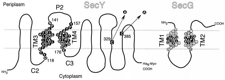
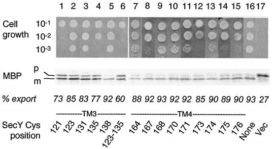
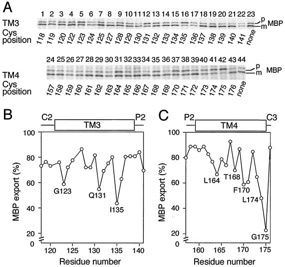
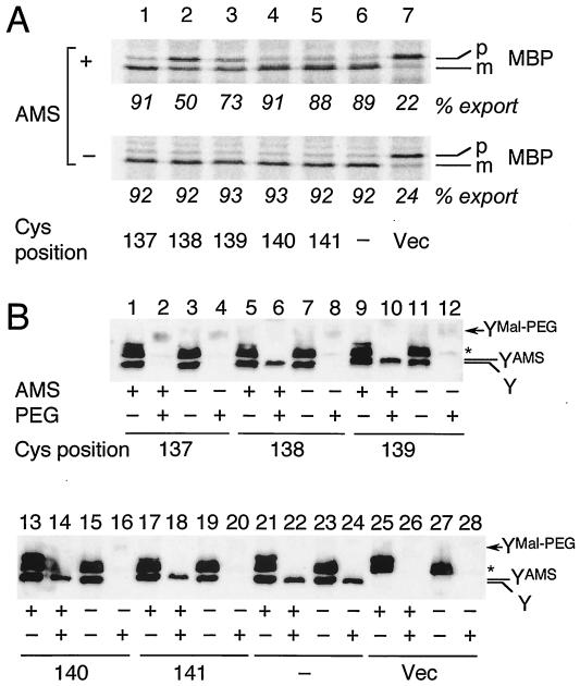
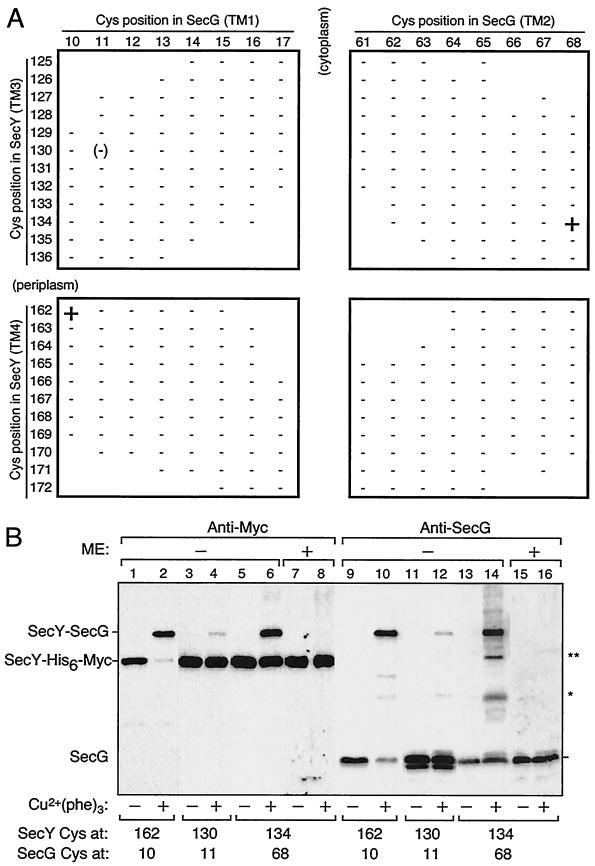
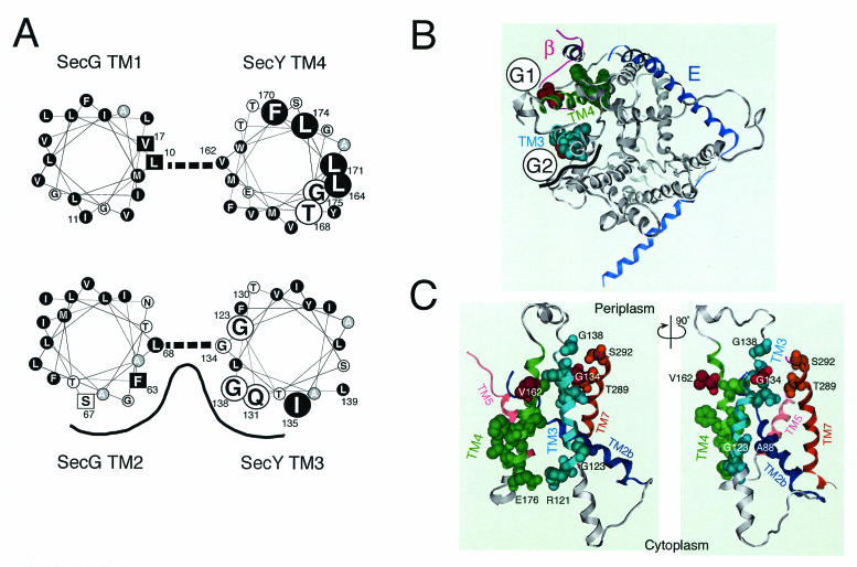
The SecYEG heterotrimeric membrane protein complex functions as a channel for protein translocation across the Escherichia coli cytoplasmic membrane. SecY is the central subunit of the SecYEG complex and contains 10 transmembrane segments (TM1 to TM10). Previous mutation studies suggested that TM3 and TM4 are particularly important for SecY function. To further characterize TM3 and TM4, we introduced a series of cysteine-scanning mutations into these segments. With one exception (an unstable product), all the mutant proteins complemented the cold-sensitive growth defect of the secY39 mutant. A combination of this secY mutation and the secG deletion resulted in synthetic lethality, and the TM3 and TM4 SecY cysteine substitution mutations were examined for their ability to complement this lethality. Although they were all positive for complementation, some of the complemented cells exhibited significant retardation of protein export. The substitution-sensitive residues in TM3 can be aligned to one side of the alpha-helix, and those in TM4 revealed a tendency for residues closer to the cytosolic side of the membrane to be more severely affected. Disulfide cross-linking experiments identified a specific contact point for TM3 and SecG TM2 as well as for TM4 and SecG TM1. Thus, although TM3 and TM4 do not contain any single residue that is absolutely required, they include functionally important helix surfaces and specific contact points with SecG. These results are discussed in light of the structural information available for the SecY complex.


Figures

FIG. 1.
FIG. 1.
SecY and SecG residues that were subjected to cysteine substitution mutation. SecY and SecG amino acid sequences are shown according to their topology models (1, 18). The two cysteine residues of SecY (black squares) were converted to alanine as indicated by the arrows. Single-cysteine mutations were then introduced into residues shown by circles with a black background. SecG residues that were converted to cysteine are similarly shown.
FIG. 2.
FIG. 2.
Abilities of single-cysteine SecY derivatives to complement the secY39 defect. Plasmids encoding single-cysteine SecY derivatives as specified by the positions (indicated by residue numbers) of substitution were introduced into strain GN31 (secY39). For growth tests (top), cells were grown in L broth at 37°C until mid-log phase and diluted serially (10−1 to 10−3 as indicated) with 0.9% NaCl; 2-μl portions of each suspension were spotted onto L-agar plates, which were incubated at 37°C for 12 h (data not shown) as well as at 20°C for 48 h (photographs). For protein export assay, the same set of bacterial cells was grown at 37°C in M9 minimal medium containing glycerol and amino acids supplemented with maltose (0.4%) and IPTG (1 mM) until early log phase. Thirty minutes after the temperature was shifted to 20°C, cells were pulse-labeled with [35S]methionine for 1 min followed by immunoprecipitation of MBP, SDS-PAGE, and phosphorimaging visualization of the labeled MBP species. The positions of the precursor (p) and mature (m) forms of MBP are indicated. The proportions (% export) of the mature form are shown below the electrophoretic patterns. The Cys position mutated in SecY is shown at the bottom of the figure. Vec, vector.
FIG. 3.
FIG. 3.
Protein export activities of the secY39 ΔsecG cells complemented with single-cysteine SecY derivatives. (A) Profiles of pulse-labeled MBP. The ΔsecG::kan mutation was introduced by P1 transduction into strain GN31 (secY39) carrying plasmid pSTD343 (lacIq) as well as a plasmid encoding the cysteine-less SecY-His6-Myc (lanes 23 and 44) or one of the single-cysteine SecY derivatives with substitutions in TM3 and TM4 at the residues indicated below the blots by residue numbers. Cells were grown at 37°C in M9 minimal medium containing glycerol, amino acids, maltose (0.4%), and IPTG (1 mM) until early log phase. The temperature of a portion of the culture was then shifted to 20°C for 30 min, and then the culture was pulse-labeled with [35S]methionine for 1 min. MBP was immunoprecipitated and separated into the precursor (p) and mature (m) forms. Labeled MBP molecules were visualized by phosphorimaging. (B and C) Graphical representations of the MBP export activities as a function of the mutation locations. The proportions of the labeled mature form of MBP (MBP export) are plotted against the mutation positions as indicated by amino acid residue numbers in TM3 (B) and TM4 (C).
FIG. 4.
FIG. 4.
(A) Effects of AMS treatment of intact cells on protein export activity. Cells of strain GN31 (secY39) carrying plasmid pSTD343 (lacIq) as well as a plasmid encoding a TM3 single-cysteine SecY derivative (lanes 1 to 5 [Cys substitution positions shown at the bottom of panel A]), cysteine-less SecY-His6-Myc (lane 6), or vector (Vec) (lane 7) were grown at 37°C in M9 minimal medium containing glycerol, amino acids, maltose (0.4%), and IPTG (1 mM) until early log phase. Each culture was divided into two portions, one of which was treated with 3 mM AMS at 37°C for 30 min (+) and not treated with AMS (−). Cells were then pulse-labeled with [35S]methionine for 30 s at 37°C and processed for MBP immunoprecipitation for visualization of labeled MBP species. The positions of the precursor (p) and mature (m) forms of MBP are indicated. The percent export values represent proportions of the mature form. (B) Verification of AMS modification by the blockage of subsequent Mal-PEG modification. Cells expressing SecY with cysteine substitutions, as indicated by the residue numbers, were grown as described above for panel A and treated with AMS as described above (+) or not treated with AMS (−). The samples were precipitated with 5% (final concentration) trichloroacetic acid and solubilized with 1% SDS supplemented with (+) or without (−) 5 mM Mal-PEG, as described in Materials and Methods. After SDS-PAGE, the SecY-His6-Myc proteins were detected by anti-Myc immunoblotting. The positions of SecY-His6-Myc (Y), its AMS-modified form (YAMS), and its Mal-PEG-modified form (YMal-PEG) are indicated. The asterisk marks a nonspecific background protein.
FIG. 5.
FIG. 5.
SecY-SecG disulfide cross-linking involving SecY TM3 and TM4. (A) SecY-SecG combinations tested. Membrane fractions prepared from cells carrying two plasmids, one expressing single-cysteine SecY-His6-Myc and the other expressing single-cysteine SecG, were subjected to Cu2+(phenanthroline)3 oxidation. The SecY-SecG combinations are indicated by the residue numbers where cysteine substitutions had been introduced. Samples were analyzed by nonreducing SDS-PAGE and immunostaining using anti-Myc and anti-SecG antibodies. Symbols: +, significant formation of a cross-linked product; −, absence of cross-linking. The 130-11 combination gave very faint cross-linked product and was scored as (−). Combinations without a plus or minus have not been tested. (B) Electrophoretic evidence for cross-linking. Membranes were incubated in the presence (+) or absence (−) of Cu2+-phenanthroline conjugate [Cu2+(phe)3] at 37°C and then electrophoresed after treatment with (+) or without (−) 5% β-mercaptoethanol (ME). Proteins were detected by anti-Myc (lanes 1 to 8) and anti-SecG (lanes 9 to 16) immunoblotting. The SecY-SecG combinations are shown at the bottom by the residues of cysteine substitutions. The identities of the products indicated by asterisks have not been established.
FIG. 6.
FIG. 6.
Important helix surfaces of TM3 and TM4. (A) Helical wheel representations and SecY-SecG contact sites. Amino acid residues in TM3 and TM4 of SecY and TM1 and TM2 of SecG are shown in helical wheel representations viewed from the periplasmic side. TM3 and TM2 as well as TM4 and TM1 are paired as shown by thick broken lines on the basis of our disulfide cross-linking results. The functionally important SecY residues identified in this study are shown by large letters. Hydrophobic amino acids are shown by circles with a black background. Alanine is shown by circles with a gray background. Residues in SecG at which suppressor mutations against the secY104 mutation have been isolated (19) are shown by squares. The proposed TM3-TM2 helix surface with relatively hydrophilic amino acids is indicated by a black line. (B) A periplasmic top view of SecY on the basis of the three-dimensional structure of the M. jannaschii SecY complex. The coordinates for the M. jannaschii SecY complex (28) were used to model E. coli SecY according to the Molecular Operating Environment program. A top view of SecY from the periplasmic side is shown in gray, with TM3 and TM4 highlighted in teal and green, respectively, and with the M. jannaschii SecE (E) (blue) and β (magenta) subunits superimposed. The functionally important TM3 and TM4 residues identified in this study are indicated by space-filled side chains, in which the residues of contact with SecG (Gly134 and Val162) are shown in red. Schematic images for the locations of the TM segments of SecG are shown by G1 and G2. The black wavy line indicates the proposed hydrophilic surface formed by SecYTM3 and SecG TM2. (C) Side views around TM3 and TM4 of SecY. A view from the putative SecG side (G1-G2 mid point in panel B) is shown on the left, whereas its ∼90°-rotated version is shown on the right. TM2b (Ser76-Pro100), TM3, TM4, TM5 (Ile187-His205), and TM7 (Asn270-Ser292) are shown in dark blue, teal, green, pink, and brown, respectively. The important TM3 and TM4 residues, including the Arg121-Glu176 pair (see the text), are space filled for their side chains, along with residues in other TMs that are discussed in the text.

Similar articles

Cited by

References

    1. Akiyama, Y., and K. Ito. 1987. Topology analysis of the SecY protein, an integral membrane protein involved in protein export in Escherichia coli. EMBO J. 6:3465-3470. - PMC - PubMed
    1. Baba, T., A. Jacq, E. Brickman, J. Beckwith, T. Taura, C. Ueguchi, Y. Akiyama, and K. Ito. 1990. Characterization of cold-sensitive secY mutants of Escherichia coli. J. Bacteriol. 172:7005-7010. - PMC - PubMed
    1. Baba, T., T. Taura, T. Shimoile, Y. Akiyama, T. Yoshihisa, and K. Ito. 1994. A cytoplasmic domain is important for the formation of a SecY-SecE translocator complex. Proc. Natl. Acad. Sci. USA 91:4539-4543. - PMC - PubMed
    1. Breyton, C., W. Haase, T. A. Rapoport, W. Kuhlbrandt, and I. Collinson. 2002. Three-dimensional structure of the bacterial protein-translocation complex SecYEG. Nature 418:662-665. - PubMed
    1. Economou, A., and W. Wickner. 1994. SecA promotes preprotein translocation by undergoing ATP-driven cycles of membrane insertion and deinsertion. Cell 78:835-843. - PubMed

Publication types

MeSH terms

LinkOut - more resources