

doi: 10.1523/JNEUROSCI.1042-04.2004.
Removal of AMPA receptors (AMPARs) from synapses is preceded by transient endocytosis of extrasynaptic AMPARs
Affiliations
- PMID: 15175386
- PMCID: PMC3309030
- DOI: 10.1523/JNEUROSCI.1042-04.2004
Item in Clipboard
Removal of AMPA receptors (AMPARs) from synapses is preceded by transient endocytosis of extrasynaptic AMPARs
J Neurosci.
.
Abstract
AMPA receptors (AMPARs) are dynamically regulated at synapses, but the time course and location of their exocytosis and endocytosis are not known. Therefore, we have used ecliptic pHluorin-tagged glutamate receptor 2 to visualize changes in AMPAR surface expression in real time. We show that synaptic and extrasynaptic AMPARs respond very differently to NMDA receptor activation; there is a rapid internalization of extrasynaptic AMPARs that precedes the delayed removal of synaptic AMPARs.
Figures
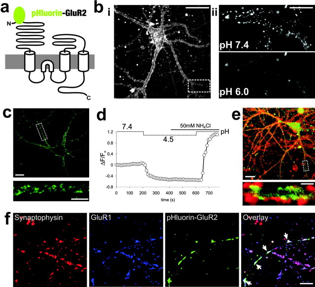
pHluorin–GluR2 reports the dynamic surface expression of AMPARs. a, Schematic of pHluorin–GluR2. b, Confocal image of typical pHluorin–GluR2 fluorescence distribution in a live, cultured hippocampal neuron. i, Fluorescence localizes predominantly to the edges of dendritic shafts. Scale bar, 20 μm. ii, The punctate, dendritic pHluorin–GluR2 fluorescence (from the dashed box in bi) seen at normal pH (7.4) is removed by washing cells with extracellular solution with reduced pH (6.0). Scale bar, 10 μm. c, Fluorescence distribution of anti-GFP surface (unpermeabilized) immunostaining from a pHluorin–GluR2-expressing neuron. Scale bar, 10 μm. The dendritic region indicated by the dashed white box in the top image is magnified in the bottom image. Scale bar, 5 μm. d, Averaged pHluorin–GluR2 fluorescence changes from dendritic regions of a typical neuron. Reducing extracellular pH causes a decrease in fluorescence as surface pHluorin–GluR2 is eclipsed. Application of NH4Cl rapidly equilibrates cellular pH levels, causing no change in fluorescence at low pH but a sharp increase at a pH of 7.4 as all the pHluorin–GluR2 fluorescence in the cell is revealed. e, Hippocampal cultures expressing pHluorin–GluR2 (green) were stained with FM4-64 (red) to visualize actively releasing presynaptic terminals. A magnified area of the dendrite indicated by the dashed box in the top image is shown in the bottom image. Most pHluorin–GluR2 fluorescent puncta are associated with presynaptic terminals (n = 4; 73 ± 3%). Scale bar, 2 μm. f, Triple labeling of synaptophysin (red), endogenous GluR1 (blue), and pHluorin–GluR2 (green) revealed that pHluorin–GluR2 is targeted to endogenous GluR1-containing synapses (white arrows in overlay image) in transfected neurons (n = 5). Scale bar, 10 μm.
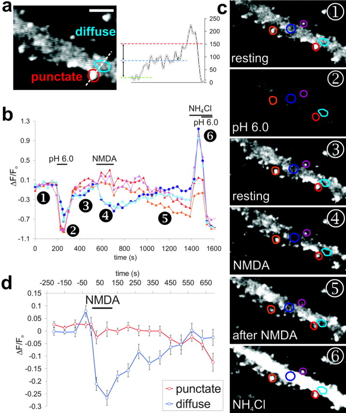
Rapid endocytosis of extrasynaptic AMPARs induced by activation of NMDARs. a, Enlarged image of dendritic pHluorin–GluR2 fluorescence labeled with typical punctate and diffuse regions. Scale bar, 4 μm. Regions for analysis were selected on the basis of relative brightness; as shown by the fluorescence profile plot (dashed white line), puncta (red line) had at least twice the fluorescence of diffuse regions (blue line). b, Fluorescence changes at individual dendritic regions shown in images in c. Lines with red tones are from typical punctate regions, and blue lines are from diffuse areas. Application of 50 μm NMDA caused a slow decrease in punctate fluorescence but a rapid and transient decrease in diffuse fluorescence. c, Images from time points corresponding to numbers on the graph. d, Binned and averaged fluorescence values from punctate (red) and diffuse (blue) regions during NMDA stimulation. Error bars show ± SEM (6 cells, 28 punctate and 27 diffuse regions).
References
-
- Banker G, Goslin K, eds (1998) Culturing nerve cells, Ed 2. Cambridge, MA: MIT.
-
- Beattie EC, Carroll RC, Yu X, Morishita W, Yasuda H, von Zastrow M, Malenka RC (2000) Regulation of AMPA receptor endocytosis by a signaling mechanism shared with LTD. Nat Neurosci 3: 1291–1300. - PubMed
-
- Blanpied TA, Scott DB, Ehlers MD (2002) Dynamics and regulation of clathrin coats at specialized endocytic zones of dendrites and spines. Neuron 36: 435–449. - PubMed
-
- Borgdorff AJ, Choquet D (2002) Regulation of AMPA receptor lateral movements. Nature 417: 649–653. - PubMed
-
- Carroll RC, Beattie EC, von Zastrow M, Malenka RC (2001) Role of AMPA receptor endocytosis in synaptic plasticity. Nat Rev Neurosci 2: 315–324. - PubMed
Publication types
MeSH terms
Substances
Grants and funding
LinkOut - more resources
Full Text Sources
Other Literature Sources
Research Materials